Fig. 34.1
Anterior view of shoulder showing the arterial supply; (1) axillary artery , (2) thoracoacromial artery, (3) subscapular artery, (4) ACHA , (5) anterolateral artery travelling superiorly on the lateral lip of the inter-tubercular groove, (6) PCHA , (7) brachial artery
At the inferior border of the teres major, the axillary artery becomes the brachial artery and continues its course distally in the arm. One of its branches, the profunda brachii, which travels through the triangular interval, partakes in anastomoses between the vasculature of the shoulder region and arm [2, 4].
34.2.1 Anterior Circumflex Humeral Artery and Its Branches
This artery has a mean diameter of 1.2 mm [3] and originates from the axillary artery at the superior edge of the pectoralis major muscle. It travels laterally under the coracobrachialis and both heads of biceps brachii, supplying these and the subscapularis as it continues towards the surgical neck. Just before reaching the surgical neck, it sends a branch to the anterior portion of the axillary recess of the joint capsule [8]. At the surgical neck it divides into four main branches. These include a descending branch to the pectoralis major insertion, a transverse branch to the periosteum of the humerus and greater tuberosity, a muscular branch to the overlying deltoid and an ascending branch known as the anterolateral artery [8]. The anterolateral artery ascends along the lateral lip of the bicipital groove, sending some penetrating branches into the lesser tuberosity along the way, and enters bone at the top of the groove [3, 7–14]. At the point where the anterolateral artery becomes intraosseous, it supplies some branches to the superior facet of the greater tuberosity and to the anterior rotator cuff interval.
The intraosseous continuation of the anterolateral artery and the principal nutrient artery of the proximal humerus is the arcuate artery , named by Laing in 1956 after its “arcuate” posteromedial course in the humeral head (Fig. 34.2) [9]. The arcuate artery supplies the lesser tuberosity, the anterior half of the greater tuberosity and most of the humeral head (Figs. 34.3 and 34.4). Given that it supplies the vast majority of the humeral head, injury or occlusion to this artery is said to increase the risk of avascular necrosis (AVN) of the humeral head [13, 15].
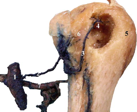
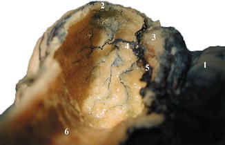
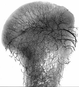
Fig. 34.2
Left proximal humerus of an ink injected specimen, lateral view, showing a continuous stretch of arteries from the axillary artery , to the anterolateral artery and finally the arcuate artery and its branches. The cortex of the greater tuberosity has been breached to follow the intraosseous vasculature: (1) axillary artery, (2) ACHA , (3) anterolateral artery, (4) arcuate artery, (5) greater tuberosity, (6) inter-tubercular groove, (7) PCHA
Fig. 34.3
Right shoulder, lateral view facing superiorly into the roof of the proximal humerus. Part of the humeral shaft and most of the cortex of the greater tuberosity have been removed to display the vasculature. The majority of the humeral head is being supplied by the arcuate artery . (1) Subscapularis, (2) greater tuberosity, (3) inter-tubercular groove, (4) arcuate artery, (5) anterolateral artery , (6) humeral shaft
Fig. 34.4
Adult proximal humerus, decalcified and with periosteum removed, showing the intraosseous arterial pattern. The largest artery to the right of the picture likely represents the arcuate artery (Copyright Henry V. Crock 1996 [16])
34.2.2 Posterior Circumflex Humeral Artery and Its Branches
The PCHA is three times bigger than its anterior counterpart [3]. After originating from the axillary artery , it travels posteriorly along the medial aspect of the surgical neck of the humerus. The PCHA usually provides branches to the inferior border of the subscapularis, the inferior aspect of the joint capsule, the lateral and long heads of the triceps muscle and the teres minor. In addition, it forms the medial and posteromedial vascular groups, a collection of vessels off the PCHA that travel along the joint capsule before ramifying and entering bone just distal to the cartilage of the humeral head (Fig. 34.5) [3, 9, 11]. These intraosseous vessels supply the surgical neck and the inferomedial aspect of the humeral head [13]. They are said to maintain the arterial supply to the humeral head in 4-part valgus impacted fractures and when a medial calcar is present [17].
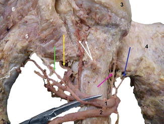
Fig. 34.5
Posteromedial aspect of a left proximal humerus showing the branches of the PCHA . The white arrows point to branches of the posteromedial artery, the pink arrow shows an anteromedial branch of the PCHA , the blue arrow is pointing to a branch supplying the subscapularis, and the branch shown by the green arrow supplies the teres minor. The yellow arrow points to the posterolateral artery : (1) PCHA , (2) subscapular artery, (3) humeral head, (4) subscapularis, (5) teres minor, (6) infraspinatus
After travelling along the medial surface of the proximal humerus and emerging through the quadrangular space , the PCHA divides into many branches. Most enter the substance of the deltoid, while one terminal branch, recently named the posterolateral artery , travels along the lateral border of the teres minor, supplying branches to the teres minor and the periosteal network over the greater tuberosity [13]. When it reaches the superior border of the teres minor, the posterolateral artery divides into a number of branches, one of which supplies the connective tissue between the teres minor and infraspinatus, while the others enter bone (Fig. 34.6a, b). Once intraosseous, these branches travel anteromedially across the greater tuberosity and humeral head, supplying the posterior half of the greater tuberosity and a posterolateral section of the humeral head (Fig. 34.7) [13].
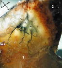
Fig. 34.6
(a) Posterolateral view of a left proximal humerus from a fresh specimen. The posterolateral artery (horizontal white arrow) highlighting its contributions to the periosteal circulation over the greater tuberosity and the connective tissue it supplies between the teres minor and infraspinatus (oblique white arrow). Full thickness rotator cuff tear presents superiorly: (1) teres minor, (2) infraspinatus, (3) greater tuberosity, (4) humeral shaft. (b) Three vascular foraminae (white arrows) on the posterolateral aspect of right proximal humerus for the osseous branches of the posterolateral artery: (1) greater tuberosity, (2) humeral head, (3) inferior facet, (4) middle facet, (5) superior facet, (6) inter-tubercular groove, (7) lesser tuberosity, (8) humeral shaft
Fig. 34.7


Looking from the inside out towards the cortex of the posterolateral aspect of the greater tuberosity. The specimen has been transilluminated to show the course of the posterolateral artery on the surface of the cortex of the greater tuberosity before it ramifies into three major branches that then become intraosseous: (1) infraspinatus, (2) teres minor
Related posts:

Stay updated, free articles. Join our Telegram channel
Full access? Get Clinical Tree
