Fig. 33.1
The suprascapular nerve runs parallel to the muscle belly of the omohyoid muscle along the posterior cervical triangle as it exits the upper trunk (Copyright K. Plancher)
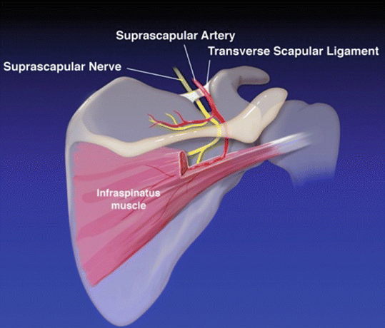
Fig. 33.2
The nerve travels along the posterior border of the clavicle to reach the superior border of the scapula. The nerve diverges from the artery to proceed under the transverse scapular ligament while the artery goes over the transverse scapular ligament diving in to the suprascapular notch (Copyright K. Plancher)
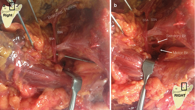
Fig. 33.3
(a, b) Suprascapular nerve coursing from the brachial plexus under the transverse ligament to enter the supraspinatus fossa and pass deep to the supraspinatus muscle. There, it gives off motor branches to the supraspinatus muscle and sensory fibers to the glenohumeral joint (Cadaveric dissections courtesy of Dr. Felix Savoie, New Orleans, LA, USA) SSN = Suprascapular Nerve; SSA = Suprascapular Artery; M = Medical; L = Lateral
The suprascapular nerve as it enters the supraspinatus fossa gives off 2 motor branches to the supraspinatus muscle belly. The nerve also gives off sensory and sympathetic branches to two-thirds of the glenohumeral joint, the coracoclavicular ligament, the coracohumeral ligament, the subacromial bursa, as well as the posterior capsule of the acromioclavicular (AC) joint [3, 11, 40]. The nerve then travels along the supraspinatus fossa heading laterally and coming within 2 cm of the posterior glenoid rim at the level of the spine of the scapula [47]. The suprascapular nerve travels laterally around the scapular spine to descend into the infraspinatus fossa only to pass under the spinoglenoid ligament (SGL), also known as the inferior transverse scapular ligament (Fig. 33.4a–c). The suprascapular nerve gives off 2–4 branches to the infraspinatus muscle belly. The suprascapular nerve is approximately 2.5 cm away from the glenoid rim and approximately 4 cm from the posterior corner of the spine of the scapula [29].
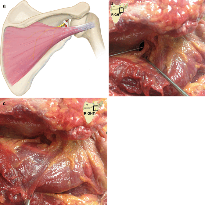
Fig. 33.4
(a) The suprascapular nerve descending into the infraspinatus fossa passing under the spinoglenoid ligament also known as the inferior transverse scapular ligament. (Copyright K. Plancher) (b, c) Cadaveric dissections of the suprascapular nerve at the spinoglenoid notch. (Cadaveric dissections courtesy of Dr. Felix Savoie, New Orleans, LA, USA) SSN = Suprascapular nerve; M = Medial; L = Lateral
33.2 Adjacent Structures and Variations
33.2.1 Suprascapular Notch
The morphology of the suprascapular notch , specifically a reduction in the height of the notch, may play a role in the development of suprascapular nerve entrapment [16]. Rengachary first classified 6 variations of the suprascapular notch [34] (Fig. 33.5). Scapula that do not have a notch, but rather a wide depression from the superior angle of the scapula to the base of the scapula, are classified as type I. A type II suprascapular notch is defined as a wide, blunted “V”-shaped notch that occupies approximately one-third of the superior border of the scapula. Symmetrical, “U”-shaped suprascapular notches, which have nearly parallel lateral margins, are classified as type III. Very small, “V”-shaped notches are classified as type IV. A type V notch is similar in shape to a type III notch with a partial ossification of the medial part of the TSL resulting in a small diameter along the superior border of the scapula. In a type VI notch, the TSL is completely ossified creating a bony foramen which is variable in size. Other classification systems of the suprascapular notch have also been described; Ticker et al. classified the suprascapular notch as either “U”- or “V”-shaped, evaluating the degree of ossification of the TSL separately, whereas, Iqbal and colleagues reported three types of notches including “U”-, “V”-, and “J”-shaped [17, 42].
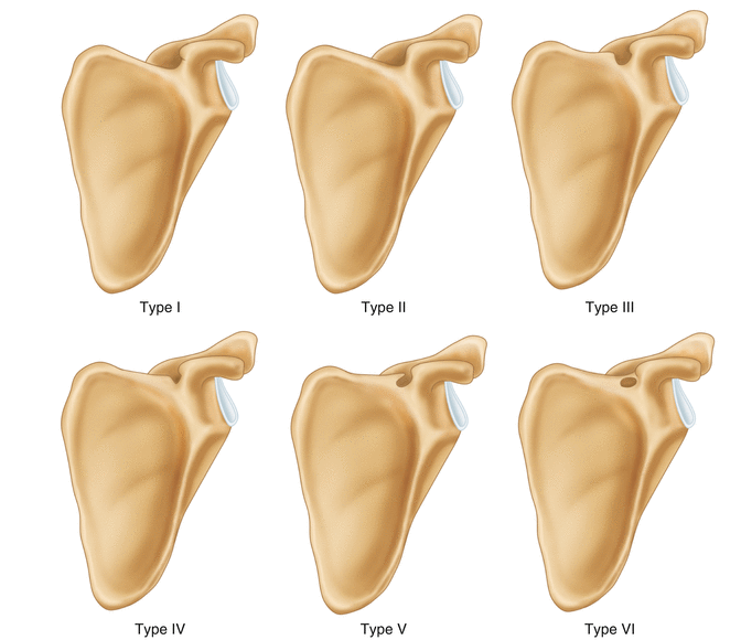
Fig. 33.5
Classification of abnormalities of the suprascapular notch by Rengachary (Adapted from Rengachary et al. [34])
33.2.2 Transverse Scapular Ligament
The TSL attaches the base of the coracoid process and the medial end of the suprascapular notch creating a foramen through which the suprascapular nerve traverses in the majority of individuals. The TSL is thin and flat, being narrower at the middle than at its insertions. Ossification of the TSL has been reported to occur in approximately 25 % of clinical cases [42]. Polguj et al. identified 3 variations of the TSL [30]. The majority of specimens exhibited either a fan-shaped ligament (54.6 %) or band-shaped ligament (41.9 %); however, a bifid ligament was also found in 3.5 % of specimens. The anterior coracoscapular ligament was present in only 51 % of specimens and contributed to a smaller area in the suprascapular notch . The presence of the anterior coracoscapular ligament may be an additional etiologic factor to consider in suprascapular nerve compression at the suprascapular notch .
Classically, the suprascapular nerve traverses under the TSL at the suprascapular notch while the suprascapular artery traverses over the TSL. In a 2014 cadaveric study, the suprascapular nerve and vein traveled below the ligament in 61.3 % of specimens [31]. Other arrangements found included (1) the suprascapular artery and vein traveling above the ligament and the nerve coursing below the ligament (17 %), (2) the suprascapular vessels and nerve all traveling below the ligament (12.3 %), and (3) other variations of the suprascapular neurovascular structures occurred in 9.4 % of specimens.
33.2.3 Spinoglenoid Ligament
The SGL is quadrangular in shape and extends from the posterior glenoid neck and posterior glenohumeral joint capsule to insert a bilaminar ligament into the scapular spine [29]. Two types of the SGL have been described: (1) type I, a thin indistinct band of tissue, and (2) type II, a well-formed ligament. The geometric shape has been described as either band-like, triangular, or irregular. Plancher et al. previously described this ligament to be present in 100 % of fresh-frozen specimens.
33.3 Pathoanatomy
Compression of the suprascapular nerve at the suprascapular notch under the TSL is the most common site of compression of the suprascapular nerve . While hypertrophy of the TSL can lead to stenosis of the suprascapular notch , the variation in the geometry of the suprascapular notch itself (see 33.2.1 Adjacent Structures and Variations – Suprascapular Notch) may also cause compression of the nerve, leading to a neuropraxia. A “V”-shaped notch with a smaller suprascapular foramen has been associated with suprascapular neuropathy [2].
33.4 Biomechanics
The SGL is a dynamic ligament. As previously mentioned, the ligament inserts onto the posterior glenohumeral joint capsule, and therefore, motion at the glenohumeral joint impacts the suprascapular nerve [29]. This insertion has larger effects upon internal rotation of the shoulder [29]. As such, when the arm moves into positions of cross-body adduction and internal rotation, the ligament tightens which can cause compression of the nerve at this distal site of the SGL [10].
On the contrary, when the arm is in positions of excessive shoulder abduction and external rotation, the medial tendinous margin of the infraspinatus and supraspinatus muscles can impinge against the lateral edge of the scapular spine, compressing the infraspinatus branch of the suprascapular nerve [34, 37]. More proximally, excessive shoulder abduction and external rotation can create an angulation against the TSL with resultant irritation to the suprascapular nerve [7, 34]. This tractioning of the nerve has been referred to as the “sling effect” because of the sharp turn the nerve takes.
Whatever the mechanism, when the nerve is subject to excessive stretch, altered nerve conduction velocity and subsequently possible clinical symptomatology can ensue. The threshold for detection of altered nerve conduction velocity due to stretch of a nerve has been shown to be at 6 % of the resting length of the nerve. Nerve stretch greater than 15 % of the resting length of the nerve leads to irreversible nerve damage [5, 41].
33.5 Diagnostic Modalities
Diagnostic modalities include imaging, diagnostic injections, and neurophysiology.
33.5.1 Imaging
While many authors have suggested that the diagnosis of suprascapular neuropathy is difficult, as it is a diagnosis of exclusion, an accurate history, detailed physical examination, and appropriate diagnostic imaging can accurately recognize this disease entity and detect any overt neoplastic disease.
Plain radiographs should always be obtained including true (Grashey) anteroposterior (AP), Y or supraspinatus outlet, axillary lateral, and Stryker notch views as well as a Zanca view to inspect the AC joint. An AP scapular view with the beam aimed 15–30° cephalad obliquely at the TSL can aid in identifying any calcifications, exostosis, or previous trauma in the form of callous formation at the notch of osseous notch variants [33, 51]. The goal of this plain film series is to detect any fractures or minute trauma to the scapula, clavicle, coracoid, or glenoid neck.
Magnetic resonance imaging (MRI) is the best imaging modality in suspected suprascapular nerve pathology because of its soft tissue resolution. Visualization of the course of the suprascapular nerve is possible with T2-weighted sagittal oblique images. Identification of soft tissue masses such as ganglion cysts has also become increasingly important when making the diagnosis of suprascapular neuropathy. The MRI will help to identify their presence, location, and size (Fig. 33.6a, b). Fritz has described the characteristic findings in asymptomatic patients with a ganglion cyst, as a homogenous signal, low T1 signal intensity with high T2 signal intensity, and rim enhancement if contrast is placed [14]. Concomitant pathologies such as labral tears which may produce secondary impingement on the suprascapular nerve , rotator cuff tendinopathy, neoplastic processes whether nerve in origin or not, and glenohumeral joint osteoarthritis can also be detected with this imaging modality. Muscle atrophy and fatty infiltration of both supraspinatus and infraspinatus, more common in chronic cases, should also be evaluated as well as the presence of muscle edema which some have suggested to be one of the earliest signs of suprascapular nerve entrapment [20] (Fig. 33.7a, b).
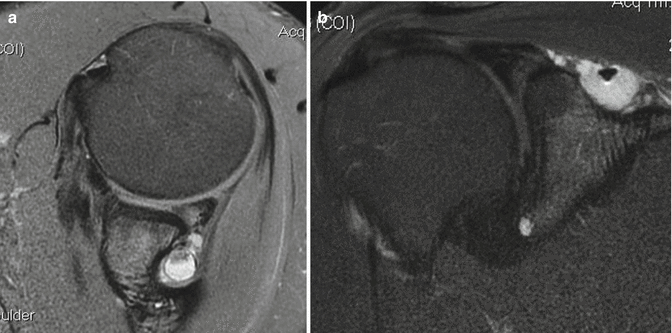
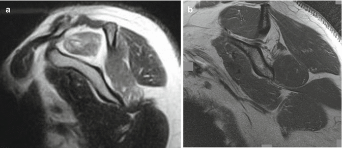
Fig. 33.6
MRI demonstrating a ganglion cyst displacing the suprascapular nerve at the spinoglenoid notch. (a) coronal, (b) axial images (Copyright K. Plancher)
Fig. 33.7
Sagittal oblique MRI demonstrating (a) supraspinatus atrophy in a young male and (b) isolated infraspinatus atrophy in a volleyball player. Note the course of the nerve in this T2-weighted image (Copyright K. Plancher)
Lastly, computed tomography (CT) and ultrasound can be valuable tools in making the diagnosis of suprascapular neuropathy. CT can detect or confirm notch variants as described by Rengachary (see 33.2.1 Adjacent Structures and Variation – Suprascapular Notch), fractures of the clavicle or scapula, and evidence of an ossified TSL [35]. Diagnostic ultrasound may also be helpful in identifying ganglion cysts in the office. In addition, ultrasound can be used to perform ultrasound-guided aspirations of a ganglion cyst.
33.5.2 Lidocaine Injection
A 1 % lidocaine anesthetic injection can be immensely helpful to accurately make the diagnosis of suprascapular nerve entrapment at either the TSL or the SGL. The authors routinely use a 25-guage, 1½-inch needle with great success. Ultrasound can be used as an adjunct to guide the needle to ensure accuracy.
When performing the diagnostic injection for compression at the TSL, it is important to understand the relationship of the artery to the nerve as previously described. The needle should be placed into the suprascapular notch from a posterosuperior approach, 3 cm medial to Nevaiser’s portal, and aiming anteriorly and aspirating first (Fig. 33.8a, b).
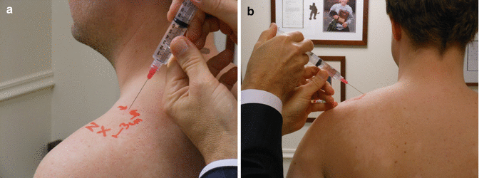
Fig. 33.8
(a) Clinical photo of a lidocaine injection to be placed at the transverse scapular ligament, 3 cm medial to Nevaiser’s portal. (b) Posterior view. Please note the angle of the needle (Copyright K. Plancher)
Injection at the spinoglenoid notch is simpler than at the TSL. When injecting into the spinoglenoid notch, the needle should be placed 4 cm medial to the posterolateral corner of the acromion (Fig. 33.9). When the injector feels the spine of the scapula, they should drop inferior to it by 1–2 cm and aspirate, and then they should easily fall into the spinoglenoid notch.
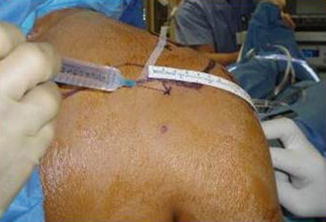
Fig. 33.9
Clinical photo of a lidocaine injection to be placed at the spinoglenoid ligament, 4 cm medial to the posterolateral corner of the acromion (Copyright K. Plancher)
Patients should be questioned regarding their pain profile immediately following the injection; pain relief can be dramatic and almost immediate. No different than when using a diagnostic injection for confirmation of impingement syndrome, the cross arm adduction test should be performed both before and after the injection to confirm the diagnosis. If previously positive, the test should now be a non-provocative maneuver. The patient may describe the absence of pain at the AC joint after this intervention, once again helping the physician in ascertaining a definite diagnosis of a suprascapular nerve compression.
A negative test, however, does not rule out the disease in those patients who have a type 4–6 notch as the ability to deliver the lidocaine is quite difficult in those situations. Patients with a negative response when there is no atrophy, a negative EMG, and no evidence of a labral tear or ganglion cyst yet present with weakness and pain require a 3-month course of nonoperative treatment before considering any type of operative intervention.
33.5.3 Electromyogram and Nerve Conduction Velocity Testing
Electrodiagnostic testing with myography and nerve conduction studies can be helpful, if positive, when there is a suspicion of the diagnosis by physical exam, imaging studies are negative (i.e., no soft tissue mass is seen), and atrophy is not present. The suprascapular nerve , as mentioned previously, is a mixed motor and sensory nerve which makes detection of a partial compression extremely difficult. Though evaluation of the sensory velocities is less useful as the sensory innervation of the suprascapular nerve is not as well defined, EMG and nerve conduction velocity testing have been shown to have 91 % accuracy in detecting nerve injury associated with muscle weakness [26, 32]. However, suprascapular nerve dysfunction can still be present with a normal EMG and nerve conduction study, making the diagnosis of suprascapular neuropathy a challenging disease entity.
Related posts:

Stay updated, free articles. Join our Telegram channel

Full access? Get Clinical Tree


