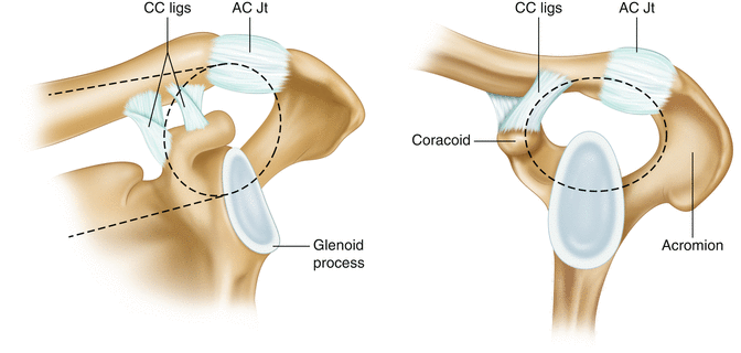
Fig. 18.1
Clavicular attachments of the conoid and trapezoid ligaments. Note the conoid attachment at the most posterior aspect of the clavicle, at the apex of curvature. These anatomic insertion points should be used for tunnel placement
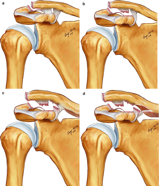
The AC joint and its stabilizing ligaments form part of the superior shoulder suspensory complex (Fig. 18.2). This is the concept of a bony and ligamentous ring that stabilizes the shoulder. Using this model, the authors of this chapter propose that rupture of the AC joint stabilizing ligaments uncouples the upper extremity from the axial skeleton as the suspensory ring is disrupted in two places. This disruption is of biomechanical significance because the AC joint transfers the cumulative forces generated by the kinetic chain originating in the lower limbs and axial skeleton when a throwing or lifting motion is initiated. Disruption of this joint will therefore disrupt this coordinated chain of loading and will alter the mechanics, power and efficiency of the desired action.
Fukuda performed a serial ligament sectioning study and reported that each ligament had a specific mode of restraint on the clavicle [3]. The AC ligaments restrain posterior displacement; the conoid ligament restrains superior but also anterior displacement and the trapezoid ligament restrains lateral displacement of the clavicle. Other authors have confirmed these findings [4]. AC joint instability can cause significant functional deficit because of deficiency of these stabilizing structures, which results in the scapula being displaced medially, inferiorly and anteriorly.
18.3 Imaging of the Unstable AC Joint
18.3.1 Plain Radiographs
The recommended radiographs for the diagnosis and quantification of AC joint injuries are a Zanca view (10–15° cephalad-tilted anterior-posterior radiograph) and axillary view to identify posterior displacement of the clavicle. Optimal exposure of the AC joint is achieved with around half the X-ray penetration required for the glenohumeral joint. Another useful view is the Stryker Notch view, which is taken with the patient supine and the arm flexed so that the hand lies on the head. The beam is directed 10° cephalad. This shows the coracoid process in profile, and should be considered if there is an AC joint dislocation with preserved coracoclavicular distance to rule out an associated coracoid fracture.
18.3.2 Stress Radiographs
Stress views can illustrate superior-inferior, anterior-posterior (AP) and medial-lateral instability of the AC joint.
AP stress views with weights strapped to the wrists have been suggested as a method to understand the degree of AC joint instability and distinguish between those dislocations with the deltotrapezial fascia intact and those without. However, these are not frequently used due to inconvenience and because the degree of instability that can be masked if the patient cannot relax the shoulder due to pain [5, 6]. The lateral stress view is a ‘scapula Y’ view in which the patient thrusts their shoulders forward to accentuate posterior-superior translation of the clavicle. This test adds little in terms of pathoanatomical understanding of the injury as compared to static views. Basamania recently described the medial stress view. This is a cross-arm adduction to demonstrate medial instability of the scapula evident when the scapula passes under the lateral clavicle [7] (Fig. 18.3). This is important from a pathoanatomical viewpoint, as chronic distal clavicle instability is a cause of scapula dyskinesia, pain and dysfunction [8].
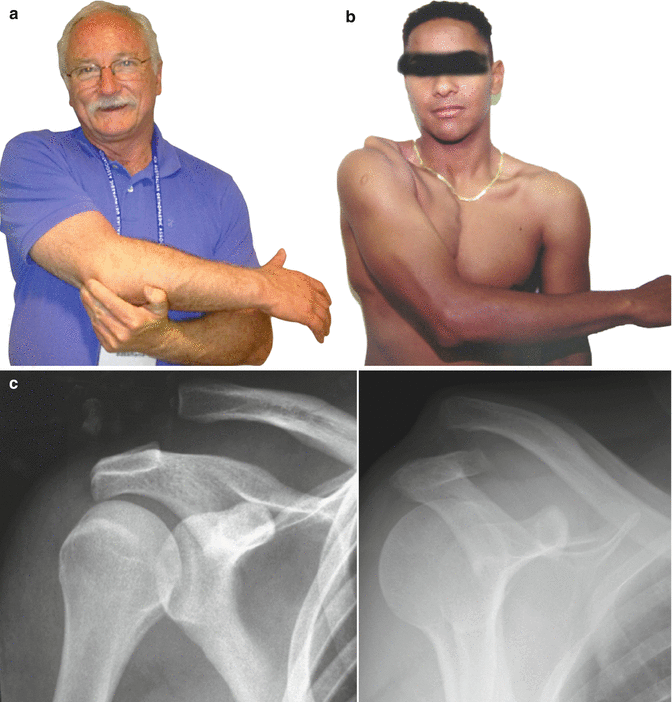

Fig. 18.3
(a–c) Cross-arm adduction provocation test. (a) The cross-arm adduction provocation test was described and demonstrated here by Carl Basamania. (b) With the arm in adduction, the prominent lateral and superior clavicle is seen. (c) Plain radiographs demonstrate that the scapula has been medialized in this Rockwood Type 5 AC joint dislocation (Image courtesy of Dr Carl Basamania, Duke University, USA)
18.3.3 Magnetic Resonance Imaging (MRI)
MRI can display the pathoanatomical structures relevant to AC joint instability especially when specific AC joint protocols are used, although interpretation can be difficult due to oedema and haematoma [9]. Moreover, AC joint instability is a dynamic entity, which is not apparent on an MRI scan. There is also evidence that MRI findings of AC joint instability do not correlate with radiographic findings or currently used classification systems [10, 11]. In our experience, we have found that the constellation of ligament injuries seen on MRI and intra-operatively is not consistent with the current classification systems.
18.3.4 4D Computed Tomography Scan (4D CT)
Advances in CT technology now allow motion capture of the skeleton in real time. We have used this to assess the unstable lateral clavicle, especially after previous surgery. It will no doubt be useful for kinematic studies, and assessment of the unstable AC joint (Video 18.1).
18.4 Pathoanatomy and Classification of AC Joint Instability
Cadenat first introduced the concept of a sequential soft tissue injury that led to progressive AC joint instability [12]. Tossy et al. [13] developed a three-stage classification, which was expanded by Rockwood in 1984 to differentiate the complete AC joint dislocations into subtypes [14]. This is the most widely used system today. These classifications were based upon clinical, radiographic and cadaveric dissections but did not include in vivo assessment or new generation imaging. Bannister [15] developed a classification system based upon stress views and intra-operative findings of the injured AC joints. In Type A the dislocation reduced, in Type B the dislocation remained unchanged and in Type C the dislocation increased. Type C were also defined as having >2 cm of coracoclavicular displacement. They documented ligamentous injuries at the time of surgery and found the coracoclavicular ligaments to be disrupted in all 21 patients they explored. The deltotrapezial fascia was sometimes torn in Type B dislocations and sometimes intact in Type C dislocations. This study highlights the discrepancy between findings at surgery and those that might be predicted.
Horn reported that the AC ligaments, the coracoclavicular ligaments and deltotrapezial fascia were torn in all nine cases. He noted that some deltoid insertion injuries were concealed under intact fascia [16]. They also reported that the meniscus of the AC joint was always avulsed from the clavicle and at least partially attached to the acromion. This study preceded any classification system so that the radiographic degree of clavicle displacement was unknown. Copeland re-emphasized the concept of sequential disruption based on his extensive personal experience and on cadaveric dissections [17]. Although not substantiated by data, he felt the injury started at the superior acromioclavicular ligaments followed by injury to the inferior acromioclavicular ligaments and periosteal stripping of the underside of the clavicle, which led to mid-substance tears of the coracoclavicular ligaments and finally deltotrapezial damage. He also stated that the meniscus always remained attached to the acromion.
Lizaur reported on the operative findings in 46 patients with ‘complete’ AC joint dislocation [18]. They found the acromioclavicular ligaments and joint capsule to be torn in all patients. The meniscus was avulsed from the clavicle in 38 patients; the coracoclavicular ligaments were torn in 40 patients and the deltotrapezial fascia torn in 43 patients. Interestingly those who had intact coracoclavicular ligaments all had a torn deltotrapezial fascia and vice versa.
All these studies address the coracoclavicular ligaments as one structure; however, they are distinct anatomic structures with different biomechanical roles and thus should not be regarded solely as one unit. Moreover, the established classification systems are not based on advanced imaging or in vivo findings, which are necessary to establish a pathoanatomical understanding of the injury.
18.5 Updated Pathoanatomy of AC Joint Instability
There has been recent recognition of the need to update the classification of AC joint instability by the International Society of Arthroscopy, Knee surgery and Orthopaedic Sports Medicine (ISAKOS) [7]. In a consensus report from ISAKOS it was proposed to subclassify Rockwood grade 3, into 3a (stable) and 3b (unstable), with this differentiation being primarily functional rather than anatomic. The 3b injuries are those with ongoing symptoms of pain and dysfunction despite a period of non-operative management. Radiographs using the Basamania cross-arm view were proposed as a method of confirming a greater degree of instability [7].
Author’s Perspective
In order to better understand the pathoanatomy of AC joint injury, we are currently prospectively assessing the advanced imaging characteristics and in vivo operative findings of acute (<4 weeks) AC joint injuries. To date, MRI scan has confirmed that in all grade 2 injuries the coracoclavicular ligaments are intact (Fig. 18.4). None of these patients underwent surgery. In Rockwood grade 3 injuries, MRI demonstrated signal change in the coracoclavicular ligaments but did not sufficiently demonstrate the details of the individual ligaments. Of the AC joint instabilities that underwent surgery, we documented that the trapezoid ligament was avulsed from the coracoid in all patients (Fig. 18.5). The conoid ligament was intact in eight out of nine grade 3 patients but often found to be lengthened (Fig. 18.6). In the grade 5 injuries the conoid was usually torn from the clavicle. The torn proximal conoid ligament often remained attached to the inferior clavicular periosteum, which was stripped medially. Figure 18.7 demonstrates a grade 5 dislocation with the typical pattern of injury. In all cases the articular meniscus remained attached to the acromion with the distal clavicle presented as a bare head with the superior acromioclavicular ligaments avulsed from the clavicle (Fig. 18.8). There was frequently a buttonhole in the deltotrapezial fascia (Fig. 18.9) and in those grade 3 injuries without a buttonhole; there was deep surface stripping from the clavicle (Fig. 18.10).
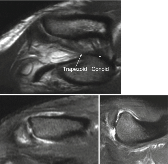
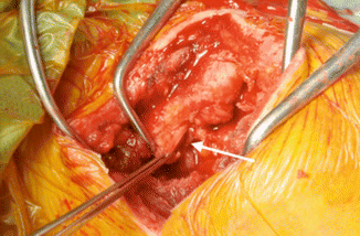
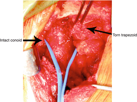
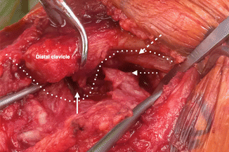
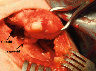
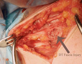
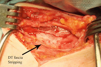

Fig. 18.4
T2 weighted MRI scans of a Rockwood type 2 AC joint injury showing intact coracoclavicular ligaments but high signal around the acromioclavicular ligaments without posterior translation of the distal clavicle (Copyright Gregory Bain)

Fig. 18.5
White arrows indicate the trapezoid ligament torn from the coracoid in—this is a typical finding (Copyright Gregory Bain)

Fig. 18.6
Grade 3 AC joint dislocation with intact conoid identified by vessel tape. Torn trapezoid from the coracoid held in forceps (Copyright Gregory Bain)

Fig. 18.7
Grade 5 AC joint dislocation. Dotted line represents the propagation of injury. Solid white arrow shows trapezoid ligament torn from the coracoid. Dotted arrow shows conoid ligament torn from the clavicle. Dashed arrow shows the inferior clavicular periosteum stripped medially (Copyright Gregory Bain)

Fig. 18.8
Bare distal clavicle with AC joint meniscus left attached to acromion (Copyright Gregory Bain)

Fig. 18.9
Buttonhole in deltotrapezial fascia (Copyright Gregory Bain)

Fig. 18.10
Concealed periosteal stripping. The superficial deltotrapezial fascia was intact (Copyright Gregory Bain)
These findings confirm that instability occurs in a progressive manner with a predictable sequential failure of the stabilizing structures. We also noted on concurrent arthroscopic examination that there was a significant incidence of superior labral tears (Fig. 18.11). This has also been noted by Imhoff who demonstrated a 14 % incidence of SLAP tears in their series with less frequent incidence of other intra-articular pathologies [19]. The reason for this may be the mechanism of injury where typically the patient falls directly onto the point of their shoulder. This drives the scapula medially causing disruption of the AC joint. In this mechanism, the humeral head will also be driven medially causing an axial loading injury to the labrum. Alternatively, it may be that patients who have concurrent labral injuries actually have a different injury mechanism with the arm above the head. This requires further investigation.
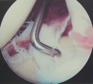

Fig. 18.11
Arthroscopic view of the glenohumeral joint in a patient with Type 3 AC joint dislocation showing the presence of a concurrent superior labral tear. This is known to be a frequent association (Copyright Gregory Bain)
We strongly feel that the coracoclavicular ligaments should be considered separately and that they, along with the acromioclavicular ligaments, each have a primary individualized function in AC joint stability. This phenomenon is evident in other joints such as the elbow, where the component parts of the medial and lateral collateral ligaments provide a specific type of restraint [20]. In an AC joint injury the ligaments fail in a predictable sequential manner. They do so because failure of one ligament results in supra-physiologic AC joint motion that further transmits the plane and magnitude of the injury to the remaining ligaments, which fail like dominos as their capacity to absorb the concentrated force is sequentially overcome. The first structures to fail are the acromioclavicular joint ligaments and capsule. The most important of these are the superior and posterior ligaments. The scapula becomes uncoupled from the axial skeleton, medializes and the clavicle externally rotates, placing the trapezoid ligament under maximal strain [3]. Trapezoid failure occurs under tension from the coracoid. There is increased coracoclavicular distance and moderate but complete superior translation of the distal clavicle, which is permitted but also limited by uncoiling and lengthening of the more posteriorly placed conoid ligament as the clavicle externally rotates (Fig. 18.12). This limit to distal clavicle displacement is apparent on radiographs, which illustrate that the degree of lateral clavicle displacement in a Rockwood grade 3 separation is consistently the same.

