Fig. 38.1
Prone position of the patient. The arm is free and is useful to paint the landmarks on the skin before the incision
The landmarks for the incisions are the acromion and the spine of the scapula that together form one continuous arch. The spine of the scapula extends obliquely across the upper four-fifths of the dorsum of the scapula and ends in a flat, smooth triangle at the medial border of the scapula. It is very easy to palpate.
38.2.2 Skin Incision
Either a horizontal or vertical incision can be chosen for posterior shoulder approach.
For horizontal approach, make a linear incision along the entire length of the scapular spine, extending to the posterior corner of the acromion. However, for vertical approach, make a vertical incision starting from the scapula spine to the axillary fold, 2 cm medial to the posterior corner of the acromion (Fig. 38.2). The central point of this incision is the same landmark as the posterior arthroscopic portal. Because the skin incision does not lie in the line of fibers of the deltoid muscle, the skin is elevated medially and laterally, dissecting between the deep fascia and overlying fat. A vertical incision at the lateral end of the scapular spine is more cosmetic but provides poor exposure of the joint.
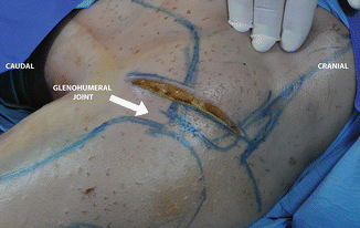

Fig. 38.2
Skin incision. A vertical incision starting from the scapula spine to axillary fold, 2 cm medial to posterior corner of acromion
Tip
To improve posterior glenohumeral joint exposure, it is useful to avoid a too lateral incision.
38.2.3 Superficial Muscle Dissection
The fascia is opened along the spine of the scapula and the muscles are exposed. The posterior part of the deltoid is now visible. The posterior deltoid muscle originates from the medial region of the scapula spine to the posterior corner of the acromion. The lateral deltoid originates from the lateral part of the acromion.
Superficial surgical dissection starts with the detachment of the posterior and small part of lateral deltoid from the scapula (Fig. 38.3). It is useful to leave some fibers of the deltoid tendon on the bone for simple reattachment at the end of the procedure. It is not simple to find the plane between the deltoid and the underlying infraspinatus muscle.
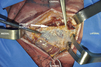

Fig. 38.3
The posterior deltoid is exposed. Detachment of the posterior deltoid from the scapula spine
Tip
To aid the dissection, it is important to identify the muscular plane that starts from the lateral end of the spine of the scapula. Once it has been found, it is not difficult to develop it if the deltoid is retracted inferiorly and the infraspinatus is exposed. Sometimes it is useful for the partial detachment of the posterior deltoid that is retracted laterally with Hohmann retractor placed between the deltoid and the humeral head (Fig. 38.4).
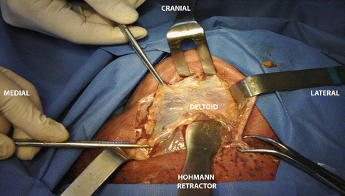

Fig. 38.4
The Hohmann retractor between the deltoid and humeral head
38.2.4 Deep Muscle Dissection
Deep dissection identifies the plane between the infraspinatus and teres minor muscles, and develops it by blunt dissection, using a finger (Fig. 38.5). This important plane can be difficult to identify. Sometimes the infraspinatus “raphe” can create confusion during dissection. The “muscular raphe” is a small fibrous band between the upper half and lower half of the infraspinatus muscle that creates a depression on the muscle. It is very dangerous to extend the dissection to the inferior border of the teres minor muscle (quadrangular space) due to the presence of the axillary nerve and the posterior circumflex humeral artery. The axillary nerve runs through the quadrangular space beneath the teres minor. Since a dissection carried out inferior to the teres minor can damage the axillary nerve (see Fig. 37.7), it is critical to identify the muscular interval between the infraspinatus and teres minor muscles and to stay within that interval. The suprascapular nerve passes around the base of the spine of the scapula as it runs from the supraspinatus fossa to the infraspinatus fossa. It is the nerve supply for both the supraspinatus and infraspinatus muscles. Gentle retraction of the infraspinatus muscle can avoid nerve apraxia after the procedure that at times may result from stretching the nerve around the unyielding lateral edge of the scapular spine.
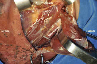

Fig. 38.5
Infraspinatus muscle and teres minor muscle are exposed. Deep dissection between infraspinatus and teres minor muscles lead to the capsular plane. Dotted line indicates the dissection plane
Danger
Damage to the posterior circumflex humeral artery in quadrangular space can create serious bleeding that is difficult to control.
Tip
Progressive internal rotation of the arm during dissection of the interval between the infraspinatus and teres minor muscle and gentle inferior retraction of teres minor can avoid damage to the axillary nerve.
38.2.5 Capsular Incision
After muscle retraction, the posteroinferior corner of the shoulder joint capsule is exposed (Fig. 38.6a, b). To explore the joint, incise it longitudinally, close to the edge of the scapula, or horizontally between the upper half and lower half of the capsule. We prefer horizontal incision in line with the interval dissection of muscles to expose the posterior glenohumeral joint (Fig. 38.7).
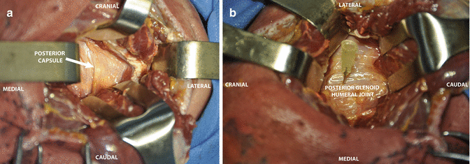
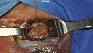

Fig. 38.6
(a) Posterior capsule is exposed. Progressive internal rotation of the arm is useful during dissection of the interval between the infraspintus and teres minor muscles. Gentle inferior retraction avoids nerve damage. (b) The needle is useful to indicate the posterior glenohumeral joint

Fig. 38.7
Posterior glenohumeral joint after horizontal capsular incision
Trick
To gain better access to the posterior aspect of the shoulder joint, it can be useful to detach the infraspinatus 1 cm from its insertion onto the greater tuberosity. Retract the muscle medially, taking great care not to damage the suprascapular nerve, which enters the undersurface of the muscle just below the spine of the scapula. If the skin incision is too latral, it is possible to have more difficulty accessing the posterior glenoid neck.
38.3 Pathoanatomy
38.3.1 Effect of Trauma
Traumatic events can create various problems in the shoulder: fractures, instability, muscle and tendon lesions. Glenohumeral instability is a relatively common condition affecting 2 % of the general population [5]. Anteroinferior glenohumeral joint instability is the most common problem after trauma. Posterior glenohumeral joint instability is far less common than anterior instability, accounting for approximately 2–10 % of all cases of shoulder instability [6]. Posterior instability of the shoulder and fracture of the glenoid sometime need arthroscopic or open treatment. Posterior approach to the shoulder is used in open procedure. Regarding posterior instability , the trauma may be macro or micro. Macrotrauma is a single event [7, 8] of an axial load to the arm while the shoulder is flexed. Microtrauma is a repetitive injury, such as straight-arm pass blocking in football or bench pressing [9, 10]. Trauma may injure the rotator cuff, labrum, and/or glenoid, contributing to posterior subluxation. Isolated sectioning of the posterior rotator cuff musculature does not increase posterior translation [11, 12].
38.3.2 Posterior Shoulder Instability and Shoulder Lesions
Posterior dislocation of the shoulder is a rare but clinically and radiologically well-defined entity. It is of interest because most are missed during initial emergency room examinations [13, 14]. There is confusion between posterior subluxation and dislocation. Posterior dislocation is an acute entity associated with trauma and with an impression defect of the humeral head (reverse Hill-Sachs lesion) [15]. Misdiagnosis can create chronic posterior dislocation. Treatment is determined by the size of the defect and the duration of the dislocation. Recurrent posterior subluxation is a distinct and separate entity, which is often not associated with trauma and one that requires completely different management, such as nonoperative treatment or posterior reconstruction of the shoulder. This condition is usually caused by epileptic fit, electric shock or trauma, such as a fall on the outstretched arm.
Many classifications of posterior shoulder instability have been described, including degree, direction, mechanism of injury, and volition [16–18]. Hawkins and McCormack [19] discussed acute posterior dislocations, chronic (fixed/locked) posterior dislocations, and recurrent posterior subluxation. Of these, recurrent posterior subluxation is the most common.
Stay updated, free articles. Join our Telegram channel

Full access? Get Clinical Tree


