Fig. 4.1
En-face view of a three-dimensional reconstruction of the right scapula demonstrating the relationship of glenoid (asterisk) to the acromion (solid arrow) and coracoid process (dotted arrow)
In the resting position, the scapula is anteriorly rotated 30° in the axial plane [1] and lies at a 60° angle to the clavicle to accommodate the thoracic rib cage [2]. In the coronal plane, the scapula is rotated cephalad approximately 3–10°, and when viewed in the sagittal plane, it is antiverted 10–20° [3].
The scapula extends from the 2nd rib to the 7th, 8th, or 9th rib at the inferior angle (Fig. 4.2). The anterior surface of the scapula is referred to as the subscapular fossa, and its concave face accommodates the posterior thoracic rib cage to form the scapulothoracic articulation. The posterior surface of the scapula contains the scapular spine, which arises from the spinal trigone on the medial scapular border to divide the posterior surface into two separate fossae: the supraspinatus and infraspinatus fossa. These fossae communicate via an area at the lateral aspect of the scapular spine referred to as the spinoglenoid notch.
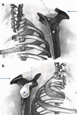
Fig. 4.2
(a) Medial view of a three-dimensional reconstruction of the right scapula demonstrating the relationship of the scapula to the thorax; note the infraspinatus fossa (large asterisk), supraglenoid fossa (small asterisk), scapular spine (dotted arrow), and acromion (solid arrow). (b) Lateral view of a three-dimensional reconstruction of the right scapula demonstrating the relationship of the scapula to the thorax; note the glenoid face (asterisk), acromion process (solid arrow), and coracoid process (dotted arrow)
The lateral aspect of the scapular spine extends beyond the scapular borders to become the acromion. The acromial angle is the angle of reflection between the scapular spine and the acromion and has a mean angle of 78° (range 64–99°) [4].
The superior scapular border contains two unique morphologies: the suprascapular notch and the coracoid process. The suprascapular notch, which is traversed by the superior transverse ligament, allows for the passage of the suprascapular nerve within the groove and into the supraspinatus fossa [5, 6]. The suprascapular artery and vein pass above the transverse ligament.
The coracoid process is a beak-shaped structure that serves as an integral attachment for muscles and ligaments acting across multiple joints in the shoulder and upper extremity facilitating both movement and stability. The coracoid serves as the attachment of the conjoined tendon, which incorporates the coracobrachialis and the short head of the biceps brachii, as well as of the pectoralis minor.
The lateral angle of the scapula narrows to form the scapular neck, which then connects the glenoid to the scapula. The most lateral aspect of the glenoid is the glenoid cavity (Fig. 4.3), which is pear-shaped, concave fossa that narrows superiorly at the anterior glenoid notch, with the inferior 2/3 of the glenoid approximating a circle [7–10]. The glenoid has two bony tubercles at the superior and inferior poles, the supraglenoid and infraglenoid tubercle, which serve as origin sites for the long head of the biceps brachii (LHB) and the long head of the triceps brachii, respectively.
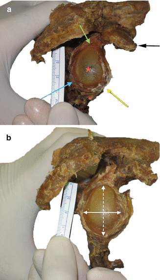
Fig. 4.3
(a) En-face view of a right cadaveric glenoid showing bare spot (large *), the projection of the coracoid (white arrow), superior labrum (green arrow), anterior inferior labrum (yellow arrow), posterior inferior labrum (blue arrow), and long head of triceps attachment (small *). (b) En-face view of a right cadaveric glenoid showing anterior to posterior distance (solid arrow) and superior to interior distance (dotted arrow)
4.2 Myology
A total of 17 individual muscles either originate or insert on the scapula, functioning across several joints in the shoulder and upper extremity, including the scapulothoracic joint, the glenohumeral joint, and the elbow.
The thoracoscapular muscles act to stabilize the scapula and provide scapulothoracic motion. These include the trapezius, the rhomboids, the levator scapulae, the serratus anterior, and the pectoralis minor. The rhomboids, levator scapulae, and trapezius muscles all originate from the cervical and thoracic spinous processes, but their insertion locations on the scapula result in distinct scapular movements.
The rhomboids insert on the superior angle and the posterior medial border of the scapula, acting to both retract (adduct) and downwardly rotate the scapula. The levator scapulae insert solely on the superior scapular angle, resulting in scapular elevation. The trapezius muscle inserts on the scapular spine and the distal third of the clavicle, opposing the rhomboids, and resulting in scapular elevation, upward/superior rotation, and abduction.
The serratus anterior muscle originates from ribs 1 through 8 or 9 and inserts on the anterior medial border and inferior angle of the scapula. The muscle acts to draw the scapula forward and to stabilize the medial scapular border to the thoracic ribcage. The serratus anterior also abducts the scapula via insertion on the inferior angle resulting in superior rotation of the glenoid cavity.
The scapulohumeral muscles primarily originate from the scapula and clavicle and insert on the humerus while acting across the glenohumeral joint to provide motion to the shoulder. The scapulohumeral muscles include the rotator cuff muscles, the teres major, the deltoids, and the coracobrachialis.
The rotator cuff consists of four muscles: the subscapularis, the supraspinatus, the infraspinatus, and the teres minor, which lie deep to the deltoid and confer both motion and stability to the glenohumeral joint. The subscapularis is the largest of the four muscles and originates within the anterior subscapular fossa, and inserts on the lesser tuberosity of the humerus where it acts to internally rotate the shoulder. The supraspinatus originates from the supraspinatus fossa on the posterior aspect of the scapula and inserts on the greater tuberosity of the humerus to abduct the shoulder in conjunction with the deltoid. The infraspinatus and teres minor work in concert to externally rotate the shoulder by inserting on the greater tuberosity. In addition to shoulder motion, the rotator cuff muscles (Table 4.1) also impart dynamic stability to the glenohumeral joint via three major mechanisms: (1) concavity compression of the humeral head into the glenoid socket, (2) coordinated muscle contraction, and (3) close association with and contribution to the glenohumeral capsule and ligaments [11].
Table 4.1
Musculature associated with the glenoid
Muscle | Origin | Insertion | Innervation | Function on scapula |
|---|---|---|---|---|
Subscapularis | Subscapularis fossa | Humeral lesser tuberosity | Subscapularis nerve | Shoulder internal rotation |
Supraspinatus | Supraspinatous fossa | Greater tuberosity of the humerus | Suprascapular nerve | Shoulder abduction, rotation |
Infraspinatus | Infraspinatous fossa | Greater tuberosity of the humerus | Suprascapular nerve | External rotation of shoulder |
Teres minor | Lateral border of scapula | Greater tuberosity of the humerus | Axillary nerve | External rotation of shoulder |
Flexion of the shoulder is accomplished primarily via the coracobrachialis muscle, which is the central of three muscles attached to the coracoid process, along with the LHB (lateral coracoid attachment) and pectoralis minor (medial coracoid attachment).
4.3 Glenoid Labrum
The glenohumeral joint is often considered to be an incongruous joint due to the size discrepancy between the humeral head and the glenoid socket [2]. This size difference is slightly reduced by the glenoid labrum, a rim of fibrocartilage tissue that effectively enlarges the glenoid cavity and reduces the inherent instability of the shoulder.
The glenoid labrum enhances the stability of the glenohumeral joint through three primary mechanisms [11]. First, the labrum deepens the concavity of the glenoid up to 9 mm in the superior-inferior direction and also doubles the antero-posterior depth to 5 mm [12]. Second, the labrum increases glenohumeral stability by increasing the surface area through which the glenoid contacts the humeral head through an arc of motion. Finally, the labrum is the site of attachment for the various glenohumeral ligaments that confer static stability to the joint [13].
Overlying and attaching to the glenoid labrum and the scapular neck is the articular capsule, which is intimately associated with the glenohumeral ligaments. The glenohumeral capsuloligamentous complex consists of the articular capsule and three articular ligaments that serve as static restraints against excessive translation of the humeral head. The anterior band of the inferior glenohumeral ligament (AIGHL) attaches to the glenoid at the antero-inferior labrum and is the primary static restraint to anterior translation when the shoulder is in an abducted and externally rotated position [11].
4.4 Vascular
The vasculature of shoulder and upper extremity arise from the subclavian artery, itself a branch of the brachiocephalic trunk on the right and the aortic arch on the left. At the medial aspect, the subclavian artery begins as a retroclavicular vessel, and as it passes between the scalene muscles at the vertex of its arch, it emerges as the axillary artery [2].
The largest branch of the axillary artery is the subscapular artery, which emerges posteriorly to the pectoralis minor, and descends medially in front of the subscapularis muscle. The artery then divides into two terminal branches, the thoracodorsal artery and the circumflex scapular artery. The thoracodorsal artery travels behind the posterior axillary fold to its course along the lateral scapular border. The circumflex scapular artery continues through the axillary space and between the two teres muscles through the omotricipital triangle.
Medially, the thyrocervical trunk branches off of the subclavian artery, giving rise to the superficial and deep transverse cervical arteries. The deep branch of the transverse cervical artery crosses the brachial plexus moving posteriorly until it reaches the superior angle of the scapula where it gives rise to a descending branch to supply the posterior muscles of the scapula. Some variants of this artery branch directly off of the subclavian artery, in which case it is known as the dorsal scapular artery.
The suprascapular artery emerges from the thyrocervical trunk just below the transverse cervical artery and courses laterally in front of the anterior scalene muscle with the phrenic nerve. The artery continues behind the clavicle towards the superior scapula border where it passes posteriorly over the superior transverse scapular ligament into the supraspinatus fossa.
Eventually, the artery enters the infraspinatus fossa via the spinoglenoid. Together, along with the scapular circumflex artery and the descending branch of the transverse cervical artery, they form the scapular anastomoses.
The thoracoacromial artery emerges from the axillary artery at the level of the upper border of the pectoralis minor. The artery pieces the clavipectoral fascia and then divides into four branches that supply the muscles of the shoulder and proximal humerus. Of these four branches, the deltoid (or humeral) branch and acromial branch are the primary blood supply to the scapulohumeral muscles.
4.5 Neurologic
The muscles of the shoulder and upper extremity are innervated by the brachial plexus, which is formed from the branches of spinal roots C5-T1. The brachial plexus is organized into roots, trunks, divisions, cords, and branches, with the branches emerging beyond the inferior border of the clavicle.
Specific to surgical scapular anatomy, the most important nerves are the suprascapular nerve and the axillary nerve. The suprascapular nerve arises from the superior trunk of the brachial plexus and passes through the suprascapular notch to enter into the supraspinatus fossa. As it passes under the superior transverse ligament, the nerve becomes most susceptible to injury via compression and sheering forces [1].
The axillary nerve runs anterior to the subscapularis, passing through the quadrangular space with the posterior circumflex humeral artery. The axillary nerve divides into three terminal branches, providing motor innervation to the deltoid and teres minor, and sensory innervation to the lateral shoulder via the superficial lateral cutaneous nerve. The anterior branch of the axillary nerve wraps around the surgical neck of the humerus approximately 3–5 mm inferior to the lateral acromion.
Related posts:

Stay updated, free articles. Join our Telegram channel

Full access? Get Clinical Tree


