Fig. 3.1
Coronal cadaveric section of the right shoulder. Note the spherical head is contained by the glenoid, labrum and rotator cuff (Section prepared by the late Pau Golano)
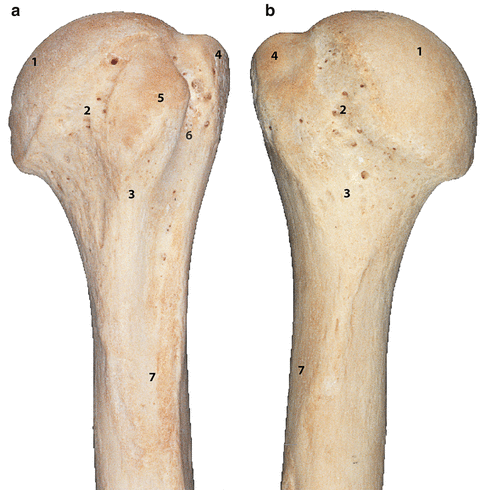
Fig. 3.2
Proximal left humerus. (a) Anterior aspect. (b) Posterior aspect. 1 head, 2 anatomical neck, 3 surgical neck, 4 greater tuberosity, 5 lesser tuberosity, intertuberous sulcus, 7 shaft, 8 radial groove (Modified with permission from Gray’s anatomy, Fig. 49.9a, b, 39th Edition, Churchill Livingstone: Elsevier, 2004, ISBN 00443071683)
The bicipital groove measures about 4 mm deep, through which the long head of the biceps tendon goes, and divides both tubercles, which form the insertion sites of the rotator cuff tendons: subscapular muscle on the minor tubercle, supraspinatus, infraspinatus and also teres minor on the major tubercle. Common findings on the humeral head are cysts, located dorsally at the insertion sites of the supraspinatus and infraspinatus tendons. In this location, humeral head cysts are not related to aging or rotator cuff tear [3]. The biceps sulcus is bridged by a band-like plate of connective tissue, the transverse humeral ligament (lig. transversum humeri), which is a continuation of the tendon of the subscapularis muscle. The only osseous fixation of this ligament lies at the medial margin of the greater tubercle of the humerus.
The surgical neck of the humerus is located distally from the major and minor tubercle. It is the region where most frequently fractures occur, due to the thin part of compact bone, which, in older age, is not anymore supported by the spongious metaphyseal bone: In older age, this is replaced by bone marrow, due to osteoporosis. The fine trabecular pattern can be visualised on the sectioned proximal humerus (Fig. 3.3).
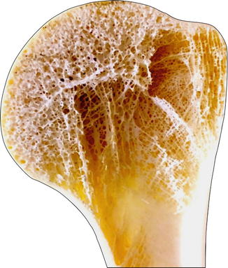
Fig. 3.3
Normal humeral head, demonstrating the trabecular pattern within the neck and head (Copyright Gregory Bain)
There exists a rotatory difference between the direction of the head of the humerus and the elbow joint. There is a retroversion of 20° between the axis of the glenohumeral joint and the axis of the elbow joint.
3.2 Vascularisation
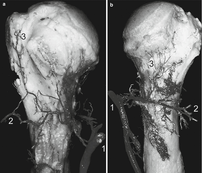
The arterial blood supply of the proximal humeral epiphysis is known to derive mainly from the anterior humeral circumflex a. (ACA) (Fig. 3.4a). However, the posterior circumflex artery (PCA) (Fig. 3.4b) is also considerably involved in the blood circulation of the bone [4]. The origin of the ACA and PCA is varied. The subchondral bone is predominantly vascularised the PCA. The apex of the head is vascularised by the ACA or the PCA equally, as is the head. The lesser tubercle derives its vascularisation mostly from the ACA, the greater tubercle from the PCA and the intertubercular groove from the ACA (Fig. 3.5a). The arcuate arteries are distributed along the metaphyseal side of the epiphyseal plate, and small branches cross the plate to reach the epiphyseal side and give numerous anastomoses to the branches of the ACA or the PCA (Fig. 3.5b). Therefore, irrespective of the choice of osteosynthesis treatment, fractures of the Neer II and 11-C AO types (fracture of the true neck) are those most vulnerable to the development of avascular necrosis. The roles of both the ACA and PCA are important and must be taken into account in evaluating the risk of necrosis after a fracture, by carefully considering the topography of the separation and the displacement of the different parts.