Fig. 30.1
Tendon of pectoralis major at its insertion onto the humerus
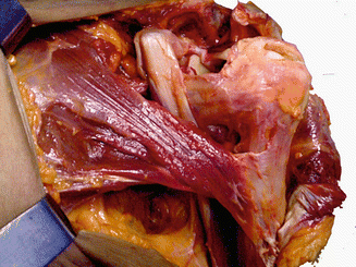
Fig. 30.2
Gross anatomy of pectoralis major muscle. Note the anterior lamina formed by the clavicular head, and the sternocostal head curling underneath and inserting progressively higher as the posterior lamina
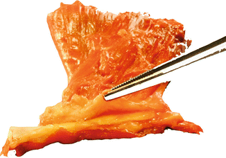
Fig. 30.3
Anatomical relationship between the pectoralis major tendon and biceps tendon
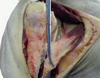
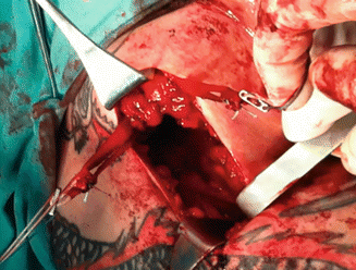
Fig. 30.4
The pectoralis major detached from its insertion demonstrating the underlying proximal biceps tendon
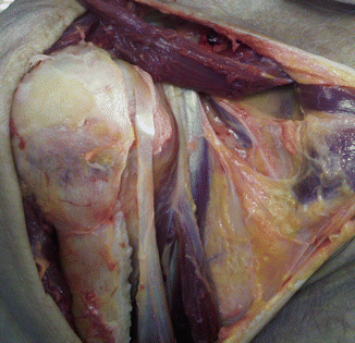
Fig. 30.5
The pectoralis major detached and reflected from its insertion demonstrating the underlying proximal biceps tendon
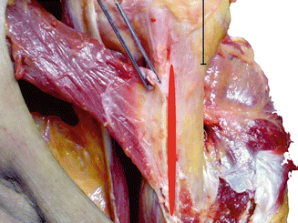
Fig. 30.6
The tendon insertion of the pectoralis major
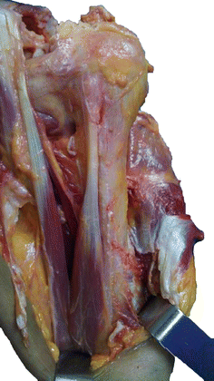
Fig. 30.7
Complete removal of pectoralis major demonstrating the underlying biceps brachii muscle
From this extensive origin the fibers converge toward their insertion; those arising from the clavicle pass obliquely downward and outwards (laterally), and are usually separated from the rest by a slight interval. Those from the sternocostal origin run upward and laterally, while the middle fibers pass horizontally. The pectoralis major tendon is bilaminar and approximately 5 cm in breadth. It is inserted into the lateral lip of the bicipital groove of the humerus (Figs 30.1, 30.2, 30.3, 30.4, 30.5, 30.6, and 30.7).
The pectoralis minor is a small triangular muscle which arises from the third, fourth, and fifth ribs under the cover of pectoralis major. It inserts via a short thick tendon onto the upper surface and medial border of the coracoid process.
30.2 Laminae
Many articles describe the pectoralis major tendon as consisting of two laminae, placed one in front of the other. They are usually blended together inferiorly.
The anterior lamina is thicker, and consists of the clavicular and the uppermost sternal fibers. They are inserted in the same order as that in which they arise. That is, the most lateral of the clavicular fibers are inserted at the upper part of the anterior lamina, while the uppermost sternal fibers pass to the lower part of the lamina. This lamina extends as low as the tendon of the deltoid muscle and joins with it (Fig. 30.2).
The posterior lamina of the tendon receives the attachment of the greater part of the sternal portion and the deep fibers from the costal cartilages. As they course upwards and laterally, they insert progressively higher into the posterior lamina of the tendon, producing the rounded appearance of the anterior axillary fold and the twisted appearance of the tendon.
The posterior lamina reaches higher on the humerus than the anterior, and from it an expansion extends over the intertubercular groove of the humerus and blends with the capsule of the shoulder joint. There is a close anatomical relationship between the pectoralis major and biceps tendons. This is consistent with our observations that in some instances following total rupture of the pectoralis major tendon, the patient also demonstrates subluxation of the long head of the biceps. It may be that the posterior lamina of the pectoralis major tendon is an important stabilizer of the biceps tendon (Fig. 30.3).
From the deepest fibers of this lamina at its insertion, an expansion is given off which lines the intertubercular groove, while from the lower border of the tendon a third expansion passes downward to the fascia of the arm.
30.3 Innervation
The pectoralis major muscle receives motor innervation by the medial and lateral pectoral nerves, so named because of their origins from the medial and lateral cords of the plexus. The medial pectoral nerve originates from the C7, C8, and T1 nerve roots from the lower trunk of the brachial plexus. It then communicates the action potential across the neuromuscular junction by releasing acetylcholine into the neuromuscular junction, inciting a proportional muscle contraction of the sternal head of the pectoralis major muscle. The second source of innervation of the pectoralis major originates from the C5 and C6 nerve roots which merge to form the upper trunk, splits off into the anterior division of the upper trunk which joins with the middle trunk to form the lateral cord. The lateral pectoral nerve branches off of the lateral cord of the brachial plexus and is distributed over the deep surface of the pectoralis major. At the neuromuscular junction, the lateral pectoral nerve provides motor input to the clavicular head of the pectoralis major.
The pectoralis minor muscle also receives innervation from both the medial and lateral pectoral nerves, from the C6 to C8 nerve roots.
The sensory feedback from the pectoralis major follows the reverse path, returning via first-order neurons to the spinal nerves at C5, C6, C8, and T1 through the anterior rami. After the synapse in the posterior horn of the spinal cord, sensory information concerning movement of the muscle, proprioception, and pressure then travels through a second-order neuron in the dorsal column medial lemniscus tract to the medulla. There, the fibers decussate to form the medial lemniscus which carries the sensory information the rest of the way to the thalamus, the “gateway to the cortex.” The thalamus diverts some sensory information to the cerebellum and the basal nuclei to complete the motor feedback loop while some sensory information ascends directly to the postcentral gyrus of the parietal lobe of the brain via third-order neurons. Sensory information for the pectoralis major is processed in the superior portion of the sensory homunculus, adjacent to the longitudinal fissure that divides the two hemispheres of the brain.
Electromyography suggests that it consists of at least six groups of muscle fibers that can be independently coordinated by the central nervous system.
30.4 Function
The pectoralis major muscle is a powerful adductor and medial rotator of the arm, and also assists in flexion of the shoulder joint. It is also responsible for keeping the arm attached to the trunk of the body, and with the upper limb fixed in abduction the muscle is a useful accessory muscle of inspiration. The two different parts are responsible for different actions. The clavicular part is in close proximity to the deltoid muscle and contributes to flexion, horizontal adduction, and inward rotation of the humerus. When at an approximate 110° angle, it contributes to adduction of the humerus. The sternocostal part is antagonistic to the clavicular part, contributing to downward and forward movement of the arm and inward rotation when accompanied by adduction. The sternal fibers can also contribute to extension, but not beyond anatomical position.
The pectoralis minor muscle is of less functional significance, but has a protective role by providing a tight band across the front of the axillary neurovascular and lymphatic contents. It assists the serratus anterior in scapular protraction and can also assist gravity in restoring the scapula to its resting position following full abduction of the arm. However, pectoralis minor has been implicated as a factor in the development of frozen shoulder.
30.5 Injuries and Imaging
Tears of the pectoralis major are uncommon, and congenital absence is rare (Figs. 30.8 and 30.9) [1–7, 10]. The most common mechanism of injury is weight lifting, in particular performing bench-press maneuvers. Most lesions are located at the musculotendinous junction and result from violent, eccentric contraction of the muscle. We have, however, also described the rupture as occurring at the transition between eccentric and concentric contractions during a bench press in athletes with stronger muscles as a result of anabolic steroids [29, 8]. A less frequent rupture site is the muscle belly, usually as a result of a direct blow [31]. Most lesions occur in male athletes, especially those practicing contact sports and weight lifting [11–27]. Women are less susceptible to these tears because of larger tendon-to-muscle diameter, greater muscular elasticity, and less energetic injuries [9]. The injury is characterized by pain in the chest wall, bruising and loss of strength of the muscle, and loss of definition of the anterior axillary fold. High-grade partial or full thickness tears often require surgery, particularly in the athletic population. Most patients are able to return to activity following surgery with high patient satisfaction and only slight reduction in strength compared to pre-injury function [28–46]. Both ultrasound and magnetic resonance imaging can confirm the diagnosis [29, 31].