Fig. 36.1
Injection of the AC joint using surface anatomy and fluoroscopic guidance techniques to localize the joint
36.1.2 Acromion
The acromion is a flat trapezoidal process that arises from the scapula spine and then projects anteriorly. Its main function is to provide an origin for the middle deltoid fibres but it also provides attachment for the acromioclavicular ligaments, capsule and the coracoacromial ligament. The posterior border is easily palpable as is the posterolateral corner, which is a key landmark for the standard posterior arthroscopic viewing portal placed just below and medial to the posterolateral corner. The posterior border of the acromion and scapula spine is best palpated by coming from inferior upward with the ‘flat of a finger’. The lateral and anterior borders of the acromion are less obvious because of the overlying deltoid but can be identified by using the ‘flat of a finger’ (Fig. 36.2). A useful surface marking is to draw a line from the ‘V’ formed by the posterior border of the clavicle and the anterior aspect of the scapula spine extending laterally over the acromion. This bisects the acromion in half and provides a guide for lateral arthroscopic portals (Fig. 36.3). The lateral border can be used to reference the position of the axillary nerve, which lies 4–7 cm inferior to it on the deep surface of the deltoid muscle [1]. The nerve should be marked on the skin in all approaches that split the deltoid and identified during the approach. The anterior lateral corner of the acromion is important for arthroscopy and open surgery. It is the site of most subacromial spurs and is an important landmark when performing an acromioplasty. This corner also marks the raphe between the anterior and middle parts of the deltoid muscle, which is a useful avascular deltoid splitting interval. Subacromial injection is a commonly performed procedure done by physicians, surgeons and allied health professionals. The subacromial bursa is located under the anterior half of the acromion and hence injections are often performed from anterior or lateral. The authors prefer injection from a posterolateral entry point because anteriorly the space between the humeral head and acromion is very small and laterally it is easy to misjudge the slope of the acromion and abut the humeral head or acromion itself. By palpating the posterolateral corner of the acromion, it is easy to guarantee entry into the subacromial space; and by directing the needle anteriorly, the bursa is very reliably injected with little discomfort or subcutaneous extravasation (Fig. 36.4).
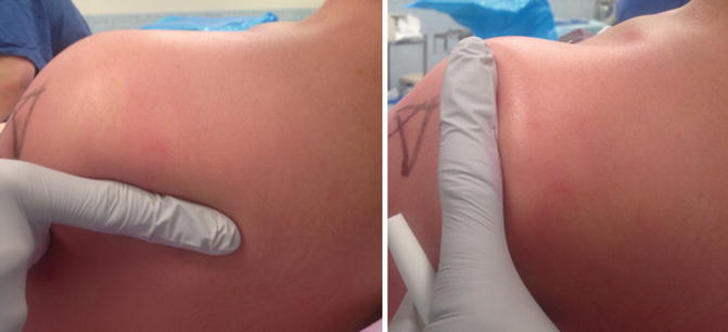
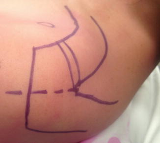
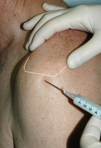
Fig. 36.2
The flat of the finger technique to identify the borders of the acromion
Fig. 36.3
Marking of the skin prior to arthroscopic surgery. Note the dotted line which serves as a guide for lateral portal placement
Fig. 36.4
Injection of the subacromial bursa from a posterolateral starting point
36.1.3 Coracoid Process
The coracoid is a key anatomic landmark in shoulder surgery. It is often visible in the thinner patient and palpable in all. It is often marked as a circle but it is worth remembering that the coracoid is hook shaped with the horizontal component being more oblong and oblique in direction. The brachial plexus, axillary artery and vein are all located on the medial side of the coracoid.
The coracoid thus serves as a safety beacon when performing surgery to the shoulder. The musculocutaneous nerve, pierces the coracobrachialis between 3 and 8 cm distal to the coracoid process [2]. The suprascapular nerve lies 1 cm medial and 2 cm posterior to the coracoid process base as it enters the suprascapular notch (Fig. 36.5).
Fig. 36.5
3D CT scan showing superior view of coracoid process and its anatomic relation to the suprascapular nerve as it passes through the suprascapular notch
Arthroscopically the coracoid is important as it provides the trajectory for entry into the glenohumeral joint from the posterior portal. The coracoid is the superficial limit of rotator interval release and can cause impingement to the upper fibres of subscapularis.
36.1.4 Glenohumeral Joint
The glenohumeral joint is deep and the joint line is not clearly palpable. However, the humeral head can be readily palpated and balloted to indicate the position of the joint. Aspiration or injection of the joint can be performed from posterior or anterior by using the surface landmarks of the coracoid and acromion. The shortest distance into the joint is via the rotator cuff interval anteriorly, which is located between the coracoid process and bicipital groove. This is located lateral to the coracoid, medial to the biceps tendon and superior to the subscapularis. The subscapularis cannot be palpated therefore staying close to the lower border of the AC joint and just lateral to the coracoid while aiming caudally by 45° will allow safe entry through the rotator interval. Alternatively, a posterior approach starting 2 cm inferior and medial to the posterolateral corner of the acromion and aiming towards the coracoid may be used (Fig. 36.6). A long needle is necessary in larger patients if this method is used.
Fig. 36.6
Glenohumeral joint injection using a posterior entry point
36.1.5 Scapula and Peri-scapular Muscles
The triangular shape of the scapula body can be identified posteriorly despite circumferential attachment of the important peri-scapular muscles. The spine of the scapula separates the supraspinatus and infraspinatus fossae. When examining a patient with shoulder pathology it is essential to identify wasting of the supraspinatus or infraspinatus, which may represent a chronic cuff tear or suprascapular nerve denervation. ‘Winging’ of the scapula is also evident when viewed from posterior and is important to note as it may be primarily or secondarily related to shoulder pathology. See Chap 29 on the anatomy of scapula winging.
36.1.6 Rotator Cuff
The cuff is difficult to feel, as it lies deep to the deltoid and to bone. The anterior superior rotator cuff insertion (supraspinatus) can be palpated on the humeral head lateral to the anterolateral aspect of the acromion and localized pain in this area can be indicative of cuff pathology. A palpable clicking can often be felt during rotation and elevation of the arm with the opposite hand palpating the cuff insertion on the humeral head. Although not described, this clicking appears to be consistent with the presence of a full thickness rotator cuff tear. The coracoacromial ligament can be felt if thickened just below the anterior edge of the acromion, which may be a source of subacromial impingement.
36.1.7 Muscles
Several large muscles drape the shoulder girdle. An understanding of their surface anatomy is useful in the localization of tenderness, for the diagnosis of pathology and for identification of inter-muscular intervals for surgical approaches to the shoulder. The deltopectoral approach is a workhorse anterior approach to the shoulder utilizing the inter-nervous plane between pectoralis major and deltoid. The coracoid process is palpated and from there the groove can be seen running obliquely across the anterior aspect of the shoulder in most individuals. Its appearance can be enhanced by externally rotating the arm to place the anterior skin under tension. By running the fingers across the anterior deltoid from lateral to medial it can be palpated as the fingers dip into the groove. In some muscular individuals the outline of the cephalic vein, which lies within the groove, can be seen through the skin. Inferior to the deltopectoral groove, the pectoralis major tendon inferior border forms the anterior axillary skin fold. Asymmetry of this fold is indicative of pectoralis major rupture in a patient with an appropriate history. The posterior axillary skin fold is defined by the latismus dorsi. Both these muscles can be made taught for inspection by pushing the hands into the hips with the shoulder abducted. The long head of biceps is a common cause of shoulder pain. It runs in the bicipital groove between the insertions of latissimus dorsi and pectoralis major on the proximal humerus. It can be palpated deeply within the deltopectoral groove especially when the shoulder is externally rotated. On the medial aspect of the upper arm the posterior aspect of the biceps muscle belly forms a groove with the anterior aspect of the triceps. This groove contains the axillary artery, basilic vein and median and ulna nerves as they exit the axilla and course distally. The anterior raphe of the deltoid can also be seen and palpated in certain patients and is an important avascular landmark utilized for dissection during deltoid splitting approaches. The axillary nerve is best evaluated by palpation over the lateral aspect of the upper arm (regimental badge area) and by resisted motor testing of all the parts of the deltoid which are supplied by discrete branches of the nerve.
Related posts:

Stay updated, free articles. Join our Telegram channel

Full access? Get Clinical Tree


