Fig. 19.1
The bony anatomy of the sternoclavicular joint including the medial clavicle, manubrium, and first rib is depicted. In addition, the sternoclavicular ligament, interclavicular ligament, costoclavicular ligament, and intra-articular disk are shown
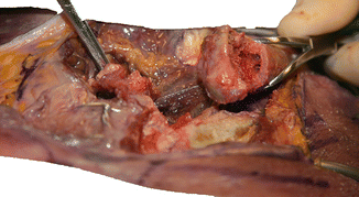
Fig. 19.2
The left medial clavicular head is shown. The medial clavicle is cut to demonstrate the bony anatomy of the medial clavicle. Note also the intra-articular disk, which is reflected from the SC joint
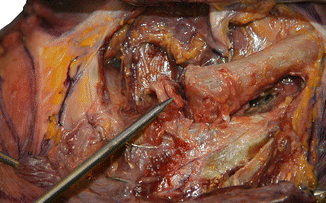
Fig. 19.3
The intra-articular disk of the left sternoclavicular joint is shown. Note that the disk in this specimen is partially degenerated. Approximately half of the medial clavicular head lies cranial to the relatively shallow clavicular notch of the manubrium. Also visualized are the sternothyroid and sternohyoid muscles posterior to the sternoclavicular joint
19.2.2 Manubrium
The manubrium is the most cranial of the three bones that constitute the sternum. It is attached to the sternal body by a synchondrosis that ossifies in middle to late adulthood [38]. The manubrium has curved, shallow clavicular notches at its superolateral borders, which articulate with both sternal heads (Fig. 19.3). These clavicular notches are covered in fibrocartilage [44]. Both the anterior and posterior sternoclavicular ligaments insert on the manubrium, while the interclavicular ligament courses along the superior portion of the manubrium (Fig. 19.1).
19.2.3 First Rib
The first rib is the broadest and shortest true rib. Anteriorly, it contains costal cartilage, which attaches to the manubrium through a synchondrosis of dense, adherent fibrocartilage [38]. This synchondrosis is located inferolaterally from the clavicular notch of the manubrium (Fig. 19.1) [38]. It represents the most inferior and lateral aspect of the sternoclavicular joint. The synchondrosis of the first rib serves as an attachment site for the costoclavicular ligament. The first ribs, along with the manubrium and first thoracic vertebrae, constitute the thoracic outlet.
19.2.4 Fibrocartilaginous Disk
The sternoclavicular joint contains an intra-articular disk, which attaches to the anterior and posterior sternoclavicular ligaments (Fig. 19.3) [50, 57]. The disk is analogous to the meniscus in the knee and divides the joint into two distinct synovial cavities [50]. It is comprised of a dense, vertically oriented, fibrocartilaginous medial sternoclavicular portion and a thinner more horizontally oriented lateral costoclavicular portion [3]. The intra-articular disk is thought to degenerate with age [50]. Van Tongel et al. [57] demonstrated that the disk ligament was incomplete in 56 % of cadaver specimens evaluated, containing centrally located holes associated with fraying of the disk and degeneration of the clavicular cartilage. The presence of an incomplete intra-articular disk ligament is likely to be related to degenerative changes rather than developmental abnormalities, because this variant was only noted in cadaver specimens greater than 75 years of age [57]. The disk is believed to enhance shock absorption, protect the articular surfaces of the SC joint, aid in rotation of the clavicle, and help prevent medial displacement of the clavicle [17, 14, 44, 55].
19.3 Ligamentous Anatomy
There is limited osseous contact between the clavicle and the manubrium; so, the stability of the sternoclavicular joint is primarily provided by the ligamentous structures [44, 55]. The stabilizing ligaments of the sternoclavicular joint include the costoclavicular ligament, interclavicular ligament, and the anterior and posterior sternoclavicular (SC) ligaments (Fig. 19.1) [3, 7, 17, 44, 50, 55, 56].
19.3.1 Costoclavicular Ligament
The costoclavicular ligament contains an anterior and a posterior fasciculus, which arises from the chondral surface of the first rib and inserts on the undersurface of the medial clavicle (Fig. 19.4) [44]. The anterior fasciculus originates on the anterior medial aspect of the chondral surface and its fibers run superolaterally, while the posterior fasciculus originates on the posterior lateral aspect and its fibers run superomedially [44]. The anterior fasciculus of the costoclavicular ligament limits both lateral translation and upward rotation of the clavicle, while the posterior fasciculus limits medial translation and downward rotation [5, 44, 55].
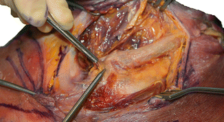

Fig. 19.4
The interclavicular ligament is shown with the use of the forceps. The probe indicates the position of the sternoclavicular joint. The fibers of the anterior sternoclavicular ligament are well visualized at the level of the joint. The anterior and posterior fasciculi of the costoclavicular ligament are seen lateral to the anterior SC ligament. Also note the internal jugular vein superolaterally from the sternoclavicular joint
19.3.2 Interclavicular Ligament
19.3.3 Capsular Ligaments
The anterior and posterior SC ligaments represent capsular thickenings, which function as the primary stabilizers of the SC joint, with the posterior ligament being the stronger of the two (Fig. 19.4) [52]. The anterior SC ligament fibers run obliquely from the medial clavicle to the sternum in a downward and medial direction, serving to prevent anterior displacement of the clavicle. The posterior SC ligament fibers cover the posterior aspect of the SC joint, preventing both anterior and posterior displacements of the clavicle [52].
19.3.4 Intra-articular Ligament
The presence of an intra-articular disk ligament is a matter of debate. Some authors argue that the intra-articular disk ligament complex passes from the dorsal and caudal clavicular head, through the SC joint, to the synchondral junction of the first rib and manubrium [3, 44, 55]. Yet others argue that the disk only inserts on the anterior and posterior capsular ligaments, which then insert onto the bone, and thus is not a ligament itself [50, 57]. Regardless, by virtue of its inferior lateral and superomedial insertions, the disk acts as a tether, preventing medial translation of the clavicular head [44].
19.4 Anatomic Structures in Close Proximity
The sternoclavicular joint is easily palpated at the base of the anterior neck. The clavicle has numerous muscular attachments including the sternohyoid, sternocleidomastoid, pectoralis major, subclavius, deltoid, and trapezius. The manubrium has attachments including the sternocleidomastoid, sternothyroid, and sternohyoid. At the sternoclavicular joint, most superficially, are the inferior fibers of the platysma and the deep cervical fascia. Deep to this lie the sternocleidomastoid muscle and the sternoclavicular joint. Pectoralis major lies inferior to the sternoclavicular joint. Immediately posterior and medial to the SC joint are the sternothyroid and sternohyoid muscles (Figs. 19.3 and 19.4). The anterior jugular vein courses anteriorly along this musculature in the region of the sternoclavicular joint. The internal jugular vein courses laterally to this musculature in the region of the SC joint, where it joins the subclavian vein to form the brachiocephalic vein (Figs. 19.4 and 19.5). The trachea exists deep and medial to these muscles. Deep and medial to the brachiocephalic vein on the right is the vagus nerve and brachiocephalic artery and on the left is the vagus nerve and common carotid artery at the level of the sternoclavicular joint (Fig. 19.6a–c). The medial supraclavicular nerve and the nerve to the subclavius innervate the SC joint. The joint receives its arterial blood supply from the branches of the internal thoracic artery and suprascapular arteries. The subclavian and external jugular veins receive venous drainage from the joint [39, 53].
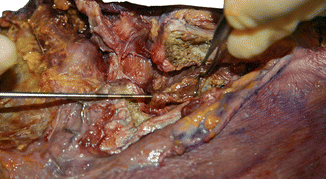
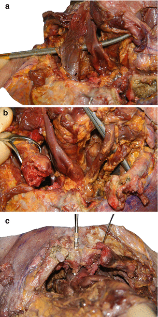

Fig. 19.5
The left brachiocephalic vein is shown posterior to the retracted clavicle at the level of the sternoclavicular joint. Also visualized is the left subclavian vein as it joins the internal jugular vein to form the brachiocephalic vein

Fig. 19.6
(a–c) The arterial structures in proximity to the sternoclavicular joint are depicted. The right common carotid artery (a) is shown deep and medial to the right internal jugular vein. The right brachiocephalic artery, left common carotid artery, and left subclavian artery (b) are shown as they branch from the arch of the aorta with the clavicles retracted and dissection of the sternothyroid and sternohyoid muscles. The trachea is also visualized deep to these structures. A drill-tip piercing the manubrium and abutting the vital arterial structures (c) is shown to emphasize the proximity of these structures to the sternoclavicular joint
19.5 Biomechanics
The sternoclavicular joint is a diarthrodial joint that has been described both as a saddle joint and as a ball-and-socket joint [42]. The osteology of the sternoclavicular joint is reciprocally concave and convex. This allows motion in coronal and sagittal planes. However, there is also a rotational component in the normal shoulder motion, which translates to the sternoclavicular joint, making it biomechanically similar to a ball-and-socket joint [28, 44]. Rockwood and others propose that the clavicle acts as a crankshaft, allowing the scapula to rotate in a 60° arc around the sternoclavicular joint [20, 45]. During the normal motion of the shoulder, the sternoclavicular joint is also capable of 30–35° of elevation and 35° of flexion and extension [45]. Motion of the sternoclavicular joint occurs mostly in the first 90° of arm elevation, with a 4° of sternoclavicular motion for every 10° of shoulder elevation. Almost no sternoclavicular motion occurs at high degrees of shoulder elevation [29].
The sternoclavicular joint is the only true diarthrodial articulation between the upper extremity and the axial skeleton in most adults. In 2.5 % of people, an articulation exists between the clavicle and the first rib [11]. However, less than half of the medial clavicle (inferior pole) articulates with the upper angle of the sternum, making this an inherently unstable joint [44].
To compensate for the lack of inherent osseous instability, the capsule and ligaments surrounding the SC joint are some of the strongest in the human body [20]. In ligament sectioning experiments using cadaveric specimens, Spencer et al. [52] found significant increases in posterior and anterior translation (107 and 42 %, respectively), which resulted from cutting the posterior capsule. Cutting the anterior capsule only produced increases in anterior translation, but to a lesser degree (26 %) than the sectioning of the posterior capsule. Cutting the costoclavicular and interclavicular ligaments had little effect on sternoclavicular joint translation. On the basis of these experiments, Spencer et al. [52] concluded that the posterior capsule is the most important restraint for posterior and anterior translation of the medial clavicle. The authors also performed a load-to-failure test on native specimens, and interestingly at a maximum load of 552 N, the failure occurred at the bone–cement interface of the testing apparatus [52]. No studies exist on what the true load-to-failure load is of the native anterior or posterior capsule.
Dynamic muscular stabilization of the sternoclavicular joint is poorly understood. Clearly, muscular stabilization plays some role, as medial excision of the clavicle in chronic instability has been reported with good clinical results [1]. In cases of atraumatic anterior subluxation, trapezius weakness has been thought to be a contributing factor. As the superior portion of the trapezius elevates the lateral clavicle, the medial clavicle is depressed, improving the stability of the sternoclavicular joint [61].
19.6 Pathoanatomy: Atraumatic Conditions
Atraumatic conditions of the sternoclavicular joint are rare, but always raise concerns, because they may be sinister, and their surgical management can theoretically be life threatening.
19.6.1 Sternocostoclavicular Hyperostosis
Sternocostoclavicular hyperostosis (SCCH) is a rare chronic inflammatory disorder of the anterior chest wall. The disease typically begins with inflammation and calcification of the sternoclavicular ligaments and sternoclavicular perichondritis, which progresses to erosive arthritis [10, 22]. With time, progressive hyperosteotic changes are seen extending to the medial clavicle, sternoclavicular joint, manubrium, first rib, and soft tissue [10, 22]. Occasionally, changes are seen in ribs two through seven [22]. Complete fusion of the sternoclavicular joints can occur after years of chronic inflammation. Biopsy specimens demonstrate nonspecific osteosclerosis with the presence of a round cell infiltrate and granulation tissue [22]. SCCH can be associated with extrasternal manifestations such as sclerosis of the axial skeleton (vertebrae, pelvis, sacroiliac joint), peripheral arthritis, and, most commonly, palmoplantar pustulosis [10, 22, 51]. The etiology of this rare disorder and the overlap between this and others that present with multifocal nonsuppurative periosteitis and hyperostosis, namely, SAPHO (synovitis, acne, pustulosis, hyperostosis, and osteitis) syndrome and chronic recurrent multifocal osteomyelitis (CRMO), is poorly understood [4]
19.6.2 Condensing Osteitis
Condensing osteitis is a rare disorder involving sclerosis and enlargement of the inferomedial clavicle, which spares the sternoclavicular joint. The condition was first described in 1974 [8], and only 40 cases have been documented in the literature [27]. The disease typically presents unilaterally in women of childbearing age without a history of trauma. Patients experience an insidious onset of pain, which may radiate to the supraclavicular fossa, and a fusiform swelling over the medial clavicle [8, 34]. Pain is exacerbated with shoulder abduction and forward flexion. The etiology is currently unknown. Some authors argue the disorder is a response to mechanical stress, while others support an infectious etiology [8, 12, 30, 34]. Radiographs and CT scans demonstrate sclerosis, minor expansion of the inferomedial clavicle, and loss of marrow space [25]. Hypointensity in the affected region of the clavicle on T1-weighted SE images and low to intermediate signal on T2-weighted SE images is noted on MRI [41]. Bone scans with technetium-99 m methylene diphosphonate or pyrophosphate demonstrates focal increased uptake in the ipsilateral medial clavicle, while both indium and gallium scans show no focal accumulation of white blood cells at the lesion, arguing against infectious etiology [34]. Imaging typically demonstrates no involvement of the sternoclavicular joint, manubrium, or first rib in this disorder. Biopsy and gross pathology show increase and thickening of the cancellous bone, periosteal reaction, inferomedial osteophyte formation, and enlargement of the clavicular head without evidence of necrosis, bony destruction, or soft tissue involvement [8, 34].
19.6.3 Friedrich’s Disease
Friedrich’s disease, or avascular necrosis of the medial clavicle, was first described in 1924 [21]. Like most conditions involving the sternoclavicular joint, Friedrich’s disease is a rare disorder with sparse literature describing the condition. The etiology is currently unknown. While the disease has been described in men, most case reports involve female patients with unilateral disease [31, 35]. Patients typically present with insidious onset of localized pain and swelling at the sternoclavicular joint. This pain tends to increase with shoulder abduction and is not associated with a direct history of trauma. Radiographs and CT scans show sclerosis that is predominantly located in the inferomedial clavicle, but may involve the entire medial clavicular head, and an irregular sternoclavicular joint with bony destruction [9, 25]. Histological evaluation demonstrates characteristic findings of avascular necrosis, namely, Haversian systems with empty lacunae and fibrotic bone marrow [19, 35]. Some argue that given the overlap between Friedrich’s disease and condensing osteitis, specifically the presence of inferomedial clavicular sclerosis, these disorders may represent the same disease with age-related radiographic differences [31].
Stay updated, free articles. Join our Telegram channel

Full access? Get Clinical Tree


