Fig. 24.1
(a) An axial non-contrast MR image demonstrates a prominent lateral extension of the coracoid. (b) Intraoperative image showing tearing of the subscapularis tendon in the same patient
24.1.2 Subacromial Impingement
Subacromial impingement Subacromial impingement has likewise been implicated in the pathogenesis of tears of the supraspinatus tendon. The Bigliani classification, previously described in this text, demonstrates the variations in morphology most commonly evaluated on a scapular-Y radiograph [1].
24.2 Rotator Cuff Tears
Well-designed, reproducible, validated classification systems that are clinically relevant are useful for understanding the spectrum of pathology encountered in the treatment of rotator cuff tears. It should be noted that some common shoulder classification systems do not demonstrate high inter-rater agreement, and that classifications do not capture all tear patterns or components [7]. Nonetheless, the use of existing systems is helpful for the purposes of understanding anatomy and severity.
Rotator cuff tears are often described by their corresponding muscle belly. It is important to remember, however, that the rotator cuff is a confluence of its four constituent tendons and tears frequently extend beyond the margins of a single tendon. Frequently, sagittal images near the cuff insertion tendon on MRI can demonstrate the segment involved in a given tear pattern.
24.2.1 Subscapularis
Subscapularis Subacromial impingement tendon pathology is easily overlooked unless careful arthroscopic examination is performed and a high index of suspicion maintained. Though no validated classification system for these tears currently exists in the literature, Fox and Romeo described a system for describing these lesions (Table. 24.1) [4].
Table 24.1
Fox and Romeo classification of subscapularis tears
Type 1: partial thickness tears |
Type 2: complete tear of upper 25 % of tendon |
Type 3: complete tear of upper 50 % of tendon |
Type 4: complete rupture of tendon |
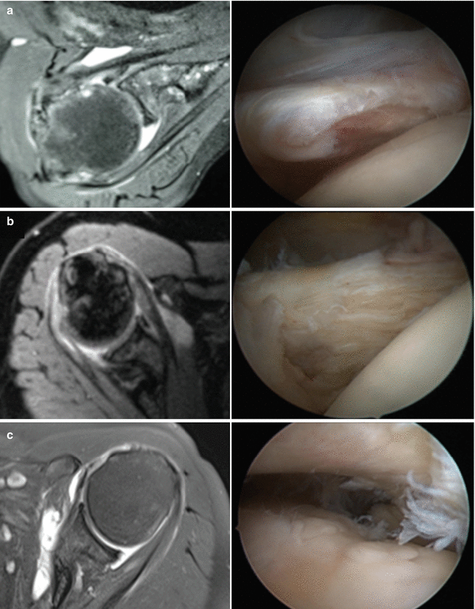
Tears of the subscapularis frequently occur at its leading, superior border. Therefore, when evaluating these lesions on MRI it is important to closely analyze the axial images immediately below the level of the coracoid as this often reveals subtle tears of the superior margin of the tendon. Figure 24.2 illustrates axial MR images and the corresponding intraoperative arthroscopic images in the same patient.


Fig. 24.2
Axial non-contrast MR image with corresponding intraoperative arthroscopic image in the same patient. (a) Type 1 lesions represent partial-thickness tears without complete tearing at any level. (b) Type 2 lesions represent complete tearing of upper 25 % of tendon. (c) Type 4 lesions represent complete rupture of the tendon
24.2.2 Partial-Thickness Tears
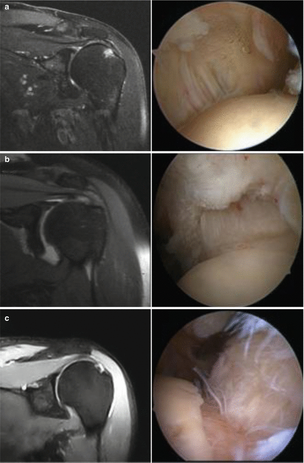
Partial-thickness of the rotator cuff may involve the articular or bursal side of the tendon. The diagnosis on is frequently more difficult than for full-thickness tears. While all imaging sequences should be closely analyzed for evidence of partial-thickness lesions, the coronal images, viewed in series, are often the most useful. The Ellman system (Fig. 24.3) of partial-thickness tears divides lesions into articular or bursal sided and determines grade by depth of tear and exposed footprint [3]. Grade 1 lesions are less than 3 mm deep, grade 2 lesions are 3–6 mm deep, and grade 3 lesions are greater than 6 mm deep. Figures 24.4 and 24.5 demonstrate articular-sided and bursal-sided partial-thickness tears, respectively, on non-contrast MRI with the associated intraoperative arthroscopic images from the same patient.


Fig. 24.4


Coronal non-contrast MR image with corresponding intraoperative arthroscopic image in the same patient. (a) Grade 1 articular-sided lesion is less than 3 mm deep. (b) Grade 2 articular-sided lesion is 3–6 mm deep. (c) Grade 3 articular-sided lesion is greater than 6 mm deep
Stay updated, free articles. Join our Telegram channel

Full access? Get Clinical Tree


