Fig. 20.1
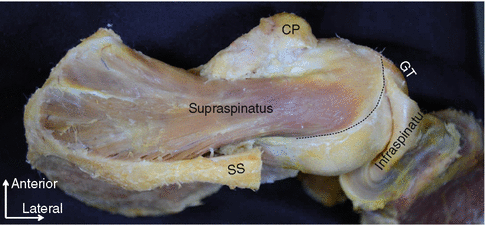
The superior view of the supraspinatus and infraspinatus (right shoulder, the acromion has been removed and reflected to anterior). Both tendons appear to mingle into one structure at the greater tuberosity (GT). SS scapular spine, CP coracoid process

Fig. 20.2
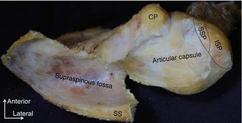
The superior view of the border between the supraspinatus and the infraspinatus (shown as the black dotted line). The infraspinatus has been detached from the scapula and the articular capsule and reflected to lateral. SS scapular spine, CP coracoid process, GT greater tuberosity
The upper surface of the greater tuberosity has been generally described as being marked by three impressions: the highest, the middle, and the lowest. However, the humeral insertion of the infraspinatus actually occupies about half of the highest impression and all of the middle impression (Fig. 20.3). The anteriormost region of the humeral insertion of the infraspinatus almost reaches the anterior margin of the highest impression of the greater tuberosity. The supraspinatus inserts into the anteromedial area of the highest impression of the greater tuberosity (Fig. 20.3). The footprint of the supraspinatus is in the shape of a right triangle, with the base lying along the articular surface. In addition to the greater tuberosity, the supraspinatus also inserts in the lesser tuberosity in one-fifth of specimens. In these specimens, the anteriormost portion of the supraspinatus tendon covers the superior part of the bicipital groove.


Fig. 20.3
The humeral side insertions of the supraspinatus and infraspinatus (the supraspinatus , SSP; the infraspinatus , ISP). Note that the articular capsule of the shoulder joint is completely separated from the supraspinatus and infraspinatus and preserved. SSP supraspinatus , ISP infraspinatus
20.2.2 Muscular and Tendinous Portions
Most of the muscle fibers of the supraspinatus , especially those of its superficial layer, run anterolaterally toward the anterior tendinous portion, while the rest of the fibers from the deep layer runs laterally toward the medial margin of the highest impression on the greater tuberosity. The supraspinatus tendon is composed of two portions: the anterior half is long and thick, and the posterior half is short and thin.
The superoanterior two-thirds of the infraspinatus is composed of a thick and long tendinous portion. A thin and short tendinous portion which occupied the rest of the infraspinatus muscle joined the thin and short tendinous portion of the teres minor .
20.2.3 Oblique and Transverse Part of the Infraspinatus
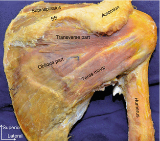
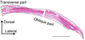
The infraspinatus is identified to be composed of oblique and transverse parts according to the direction of muscle fibers (Fig. 20.4) [6]. The oblique part is a fan-shaped muscle bundle and originates from the infraspinatus fossa running superolaterally. The transverse part originates from the inferior surface of the spine and passes laterally; it then attached to the oblique part of the middle portion of the tendinous part. Both parts are connected to each other at the superior area of the muscular portions; however, in the distal tendinous portions, they can be clearly separated. Although the oblique part is to the greater tuberosity, the transverse part does not reach the tuberosity (Fig. 20.5). The transverse part adjoined the posterior surface of the middle area of the tendinous portion of the oblique part. It is suggested that significant strength from the oblique part of the infraspinatus can focus more anteriorly and contribute to the shoulder abduction; on the other hand, the transverse part may play only a supportive role in the infraspinatus function and stabilize the tendinous portion of the oblique part during the shoulder motion from above.


Fig. 20.4
The posterior view of the right shoulder. The transverse part of the infraspinatus is shown to attach to the oblique part, SS scapular spine

Fig. 20.5
Histological section of the distal part of the infraspinatus stained by hematoxylin-eosin stain. The longitudinal section through the distal part of the infraspinatus is shown. The transverse part is shown as the dorsal dotted area. The oblique part is shown as ventral dotted area. Scale bar 10 mm
Origins of the innervating branch of the suprascapular nerve to the transverse part of the infraspinatus are variable. Branches arise from the branch to the supraspinatus muscle and/or from the main trunk of the suprascapular nerve after branching off the branches to the supraspinatus muscle (Fig. 20.6). No branch is found to pierce the transverse part to innervate the oblique part and vice versa. Although the transverse part is a part of the infraspinatus , according to its innervation, the transverse part might be closely more related to the supraspinatus .


Fig. 20.6


Schematic illustrations represented origins of the branch to the transverse part of the infraspinatus . (a) Branches arise from branches to the supraspinatus muscle. (b) Branches arise from branches to the infraspinatus muscle. (c) Branches arise from branches to both muscles. SSN suprascapular nerve, SSP supraspinatus , ISP infraspinatus
Stay updated, free articles. Join our Telegram channel

Full access? Get Clinical Tree


