Periprosthetic fractures
Clinical presentation and initial evaluation of periprosthetic fractures about the hip and knee
Incidence, classification and treatment algorithms
Technical principles and considerations
Results and reported complications
INTRODUCTION
Periprosthetic fractures about the hip and knee are a cause of significant morbidity in the geriatric population. These fractures often occur in the setting of poor bone stock secondary to osteoporosis, osteolysis or both, thereby adding a further level of complexity to already challenging reconstructions. The incidence of these fractures is increasing in parallel with the continued rise in demand for primary and revision total joint arthroplasty.1 This chapter discusses the presentation, work-up and classification of periprosthetic fractures about the hip and knee. Technical considerations and principles in the treatment of these injuries are highlighted and a review of the reported results and complications following surgical treatment of periprosthetic fractures is presented.
CLINICAL PRESENTATION AND INITIAL EVALUATION OF PERIPROSTHETIC FRACTURES ABOUT THE HIP AND KNEE
Periprosthetic fractures occurring about a total hip or knee arthroplasty can occur intraoperatively during component placement or remotely from surgery in the setting of either low or high energy trauma. This section focuses on those occurring remotely from surgery, but the technical principles applied to the surgical treatment of these fractures are universally applicable.
Evaluation of the patient with a periprosthetic fracture in the setting of high energy trauma must initially follow the guidelines of appropriate resuscitation with emphasis being placed on the airway, breathing and circulation of the patent. A thorough secondary survey is important in ruling out associated injuries. Fractures resulting from low energy injuries such as ground level falls should initiate an appropriate work-up in a geriatric patient including adequate head and neck imaging when warranted. Additionally, other organic reasons for a non-mechanical fall such as a myocardial infarction or stroke must be ruled out. Patients may benefit from a provisional reduction and splint/brace application or traction during this period of medical stabilization.
Once the appropriate initial medical work-up and resuscitation has been completed and the patient is clinically stable, important details regarding the history of the affected implant must be collected. Sometimes these details need to be gathered from family members or previous medical providers if the patient is unable to provide them. Key components of this history include antecedent pain in the affected joint, a history of infection or wound healing complications and previous operative reports that include implant types and sizes. If details in the history raise concern for the possibility of a coexistent periprosthetic infection, the joint should be aspirated to obtain a synovial fluid nucleated cell count, differential and cultures prior to reconstructive efforts.2 Intraoperative issue biopsy in conjunction with aspiration results may also improve sensitivity and specificity for the diagnosis of a concurrent periprosthetic infection.3 Serum markers for inflammation in the setting of fracture may be falsely elevated and should not be relied upon solely in dictating treatment.4
Appropriate imaging is paramount in accurately classifying periprosthetic fractures to determine the appropriate treatment strategy. Radiographs of the hip and entire femur should be obtained for all periprosthetic hip fractures around a femoral component. The presence of a long-stemmed total knee below such a fracture will greatly affect the reconstruction options available to the treating surgeon and should be anticipated. Conversely, femur views are also important in detecting a total hip arthroplasty (THA) above a periprosthetic fracture occurring around the femoral component of a total knee arthroplasty. All radiographs should be scrutinized for evidence of prosthetic loosening indicated by either a shift in prosthetic position, concentric radiolucent lines, fractured cement mantles or prosthetic subsidence. Judet views should be obtained for periprosthetic acetabular fractures to determine the columnar integrity and rule out a pelvic discontinuity. Computed tomography with three-dimensional reformatting is also useful in these instances and plays an important role in evaluating the extent of pre-existing osteolysis in the pelvis when present.
The status of the unaffected components must also be scrutinized radiographically. For the hip this is usually the acetabular component and liner. Surgical intervention to treat a periprosthetic fracture around a femoral component often affords a surgeon with a valuable opportunity to bone-graft areas of osteolysis behind an otherwise stable acetabular component, change an eccentrically worn liner or increase the head size of the reconstructed femur and potentially enhance postoperative stability. For periprosthetic fractures about the knee, the fate of two components are more closely linked, as revision of one component will often dictate the revision of the other so that the articulation and level of constraint match.
The majority of periprosthetic fractures occur in geriatric patients with osteoporosis. As such, the comprehensive care of these elderly patients should include an evaluation of their bone health. This usually involves obtaining vitamin D levels, referring the patient for bone densitometry, and potentially evaluation by a metabolic bone specialist. Often, developing a standing partnership between surgeons, endocrinologists or other primary care physicians is critical in reliably establishing a bisphosphonate treatment program for these patients to prevent future fragility fractures.
INCIDENCE, CLASSIFICATION AND TREATMENT ALGORITHMS
Hip
Periprosthetic fractures of the hip can occur in both the femur and acetabulum. Fractures of the femur occur more commonly in a revision setting and when uncemented stems are used. The risk of intraoperative fractures reported from the Mayo Clinic was 0.3% for cemented and 5.4% for uncemented stems.5 This study and others have estimated the cumulative incidence of postoperative periprosthetic femoral fractures to be 1% and 4% for primary and revision THAs, respectively.5,6 Intraoperative fractures of press-fit acetabular components have been reported with an incidence of 0.4%, and have been noted more commonly in ellipsoidal cup designs.7 Periprosthetic acetabular fractures can also occur as late sequelae of osteolysis behind acetabular components, or ground level falls in osteoporotic bone.8 Recent studies have indicated that the number of periprosthetic fractures is increasing, which places a significant burden on the healthcare system secondary to the high cost associated with the care of these complex injuries.
Multiple classification schemes exist for periprosthetic fractures about a THA; however, the most widely used and accepted is the Vancouver classification system.9,10 This system focuses on fractures occurring around the femoral component and is particularly useful in that it guides treatment strategies. It divides fractures based on location, with Vancouver A fractures occurring at a trochanteric level, Vancouver B fractures occurring about the indwelling prosthesis (usually involving the tip of the stem) and Vancouver C stems occurring distal to the stem. There is further subclassification of Vancouver A fractures depending on whether the greater trochanter (AG) or lesser trochanter is involved (AL). Vancouver B fractures are also subclassified according to the stability of the indwelling femoral component and surrounding bone quality. Vancouver B1 fractures occur around a stable femoral component, while Vancouver B2 fractures have either directly resulted from component loosening or have occurred around an already loose component. Vancouver B3 fractures involve a loose component in the setting of poor bone stock (osteoporosis, extensive osteolysis, etc).
The treatment of Vancouver A fractures (both AG and AL) usually involves a period of protected weight bearing with an assistive device. If significant displacement of an AG fracture exists, resulting in compromise of abductor function, a symptomatic limp or instability, then open reduction internal fixation (ORIF) may be indicated. Isolated periprosthetic fractures of the lesser trochanter are rare, and careful evaluation of imaging is required to ensure that other osseous involvement is not present. The majority of Vancouver A fractures occur through areas of advanced osteolysis, prompting evaluation of the bearing couple and revision when indicated to eliminate the potential particulate debris generator.
Vancouver B fracture treatment depends primarily on the status of femoral component fixation. Fractures around stable femoral implants (B1s) can be treated with open reduction and internal fixation. The use of locking plates to create balanced fixation proximal and distal to the femoral component is particularly important when performing these fracture repairs. Unicortical locking screws and judicial use of cables around the proximal segment are often required to achieve fixation. Some plates with offset screw trajectories allow for bicortical screw placement around the stem proximally which is biomechanically favourable and should be sought.11 This is more easily achieved if the indwelling implant has a dual taper design (Figure 17.1a through e).
Fractures occurring around loose stems or resulting in stem dislodgement (B2s and B3s) require advanced reconstructive efforts, combining ORIF of the femur and revision THA. Revision to long-stemmed components which bypass the fracture by two cortical diameters is recommended. Fracture patterns that involve the trochanter or fractures with significant comminution may also benefit from augmentation with a cable plate or locking plate around the revision stem. If fracture length precludes bypassing it with a stem, then plating around the revision stem with a distal femoral locking plate is also recommended.
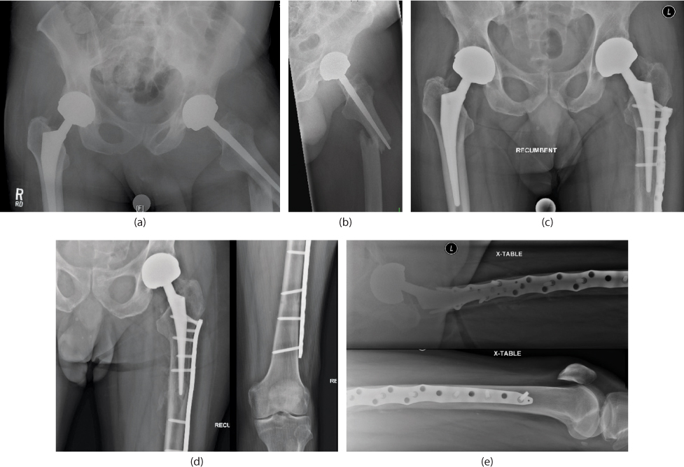
Figure 17.1 Preoperative (a,b) and postoperative (c–e) radiographs demonstrating locking plate fixation of a Vancouver B1 periprosthetic fracture. Note the screw trajectory allows for bicortical fixation proximal and distal to the tip of the stem, facilitating balanced fixation and improvement of mechanical strength.
The choice of revision stem type is dependent on the available bone stock (Figure 17.2a through d). Diaphyseal fixation should be relied on in all instances; however, if 4 cm of press fit distal to the fracture cannot be reliably achieved or the bone stock is otherwise compromised (B3), a tapered-fluted stem might be preferred.12,13 (Figure 17.3a through e). In cases where proximal bone stock is completely unsupportive of prosthetic revision, proximal femoral replacement may be indicated (Figure 17.4a through e).
As treatment of Vancouver B fractures is dictated by component stability, accurate determination of component fixation status is of paramount importance when treating these injuries.14 Preoperative imaging needs to be carefully scrutinized for signs of loosening. For cemented stems this includes fractures of the cement mantle, concentric radiolucent lines around the component or cement mantle or a shift in component position noted on serial films. For uncemented stems this includes concentric radiolucent lines, component migration or subsidence and a pedestal sign. Even in the absence of these radiographic signs, intraoperative assessment and testing of the stem should be performed, as failure to recognize a loose stem can lead to early failure of internal fixation and reconstructive efforts.
Vancouver C fractures should be treated with open reduction and internal fixation. The indwelling stem proximal to the fracture limits the role of intramedullary fixation. Good results with fixation constructs employing locking plates with balanced fixation between distal and proximal fracture segments have been observed.15 Mechanical stability is optimized with long constructs that bypass the end of the proximal stem.16 One potential caveat arises in a patient who sustains a Vancouver C fracture below a symptomatic loose stem. In this instance a revision arthroplasty with a long-stemmed component can be considered in conjunction with ORIF of the fracture, especially if the fracture is proximal enough to allow bypassing with the revision stem.
Knee
Periprosthetic fractures occurring around a TKA can occur in the femur, tibia or patella, with the femur being most commonly affected. These fractures occur intraoperatively and postoperatively, and are more commonly encountered during or following revision TKA. The reported incidence of periprosthetic fractures about a TKA is between 0.3% and 2.5%17 and they have been reported to account for 4.7% of revision TKA volume.18
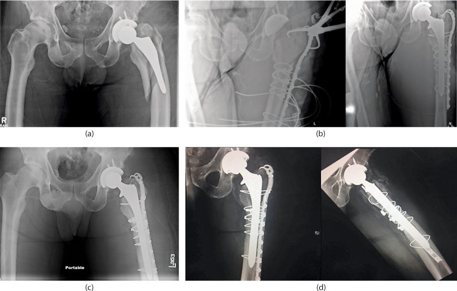
Figure 17.2 Preoperative (a), intraoperative (b) and postoperative (c,d) radiographs of a Vancouver B2 fracture treated with open reduction internal fixation (ORIF) of the femoral fracture and revision total hip arthroplasty (THA) to a monolithic, fully-porous, cylindrical stem. Note the intraoperative films which demonstrate anatomic reconstruction of the femur prior to revision stem preparation.
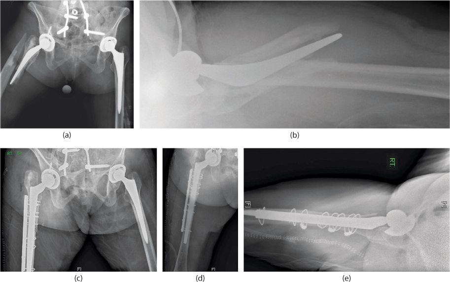
Figure 17.3 Preoperative (a,b) and postoperative (c–e) radiographs of a Vancouver B3 periprosthetic fracture which occurred around a cemented stem that was revised to a fluted, tapered, modular stem. The distal segment was prepared, the stem potted, the proximal stem assembled and the proximal fragments then cabled around the proximal body of the stem to facilitate their reduction. The acetabular liner was also changed so that a 36 mm head could be utilized to maximize stability.
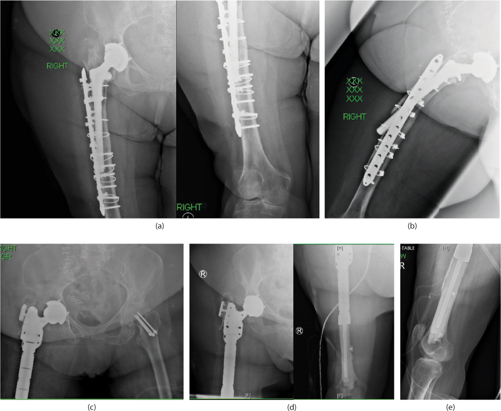
Figure 17.4 Preoperative (a,b) and postoperative (c–e) radiographs of a patient who had failed ORIF of a periprosthetic B3 fracture twice at outside institutions. The proximal segment had malunited with subsequent stem subsidence and anterior cortex perforation. The patient was subsequently revised to a proximal femoral replacement.
Related posts:

Stay updated, free articles. Join our Telegram channel

Full access? Get Clinical Tree


