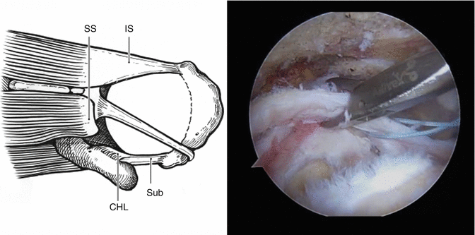
Fig. 25.1
Top row: Left shoulder, posterior viewing portal, 70° arthroscopic view of glenohumeral joint. (a) The subscapularis tendon (SSc) is torn from the lesser tuberosity (LT), but not retracted. (b) The hemorrhagic and edematous bursal surface of the tendon (IL, impingement lesion) shows evidence of extrinsic impingement at the coracoid tip (CT), which is also affected. (c) After coracoplasty and debridement, there is an adequate subcoracoid space. Left shoulder, posterior viewing portal, 30° arthroscopic view of subacromial space. (d) The sharp and worn edge of the coracoacromial ligament (CAL) and lateral acromion against a high-grade bursal-sided supraspinatus tear (BT). (e) Burr against a down sloping lateral acromial osteophyte. (f) Adequate subacromial space after decompression including beveling of lateral acromion (A)
Typically, extrinsic impingement results in abrasive lesions between the opposing structures (Fig. 25.1b, d). However, we also believe that extrinsic impingement creates high tensile forces in the articular tendon fibers (the roller-ringer effect) and can initiate either articular-sided (Fig. 25.1a) or bursal-sided tears [5]. In addition to improving visualization and working space, a thorough subacromial decompression in conjunction with rotator cuff repair has been associated with a lower rate of reoperation and re-repair even at short-term follow-up in high-level studies [6, 7]. Even in massive rotator cuff tears, where we preserve the coracoacromial ligament to prevent anterosuperior escape of the humeral head if the cuff fails to heal, we still take care to expose and treat all other potential sources of extrinsic impingement.
25.3 Full-Thickness Rotator Cuff Tears
Full-thickness rotator cuff tears present at operation with varying degrees of retraction, scarring, and delamination. The surgeon must differentiate rotator cuff tissue from “bursal leaders,” which are thickened, synovialized bands of bursal scar tissue that have an appearance similar to a chronically torn rotator cuff edge (Fig. 25.2) [8]. Bursal leaders insert into the internal deltoid fascia, whereas the intact cuff edge inserts onto the tuberosity. The surgeon must develop the plane between these two tissue edges (Videos 25.2 and 25.3) in order to correctly identify and repair the torn cuff. When cuff delamination occurs, typically only in large and massive tears, each layer should be assessed individually to determine the best repair construct. Not uncommonly, the superficial layer can be repaired with a double-row construct, while the deep layer may only be amenable to single row repair in order to avoid over tensioning.
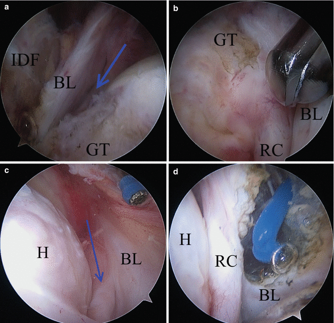

Fig. 25.2
Top row: Right shoulder. Bursal leaders as seen in a massive posterosuperior rotator cuff tear . (a) Lateral portal, 70° arthroscopic view. A deceptively thick bursal leader (BL) travels past the greater tuberosity (GT) to insert into the internal deltoid fascia (IDF), while the blue arrow identifies the intact teres minor tendon . (b) Switching the 70° scope to the posterior portal gives an enhanced view of the plane between intact rotator cuff (RC) and BL. Bottom row: Left shoulder. (c) Lateral portal, 70° arthroscopic view. A sheet-like bursal leader inserts into the internal deltoid fascia, while the rotator cuff (blue arrow) inserts into the tuberosity. (d) The bursal leader has been debrided, and the plane between the deltoid and cuff has been re-established (eg H humeral head)
Full-thickness, posterosuperior rotator cuff tears retract in several consistent patterns. The senior author (SSB) developed the geometric classification system for these supraspinatus, infraspinatus, and teres minor cuff tears based on these patterns (Table 25.1) [9]. The surgeon assesses both the size and mobility of the tear edges at the time of surgery using a tendon grasping instrument. The classification system is useful for both diagnosis and enables the creation of a treatment algorithm (Fig. 25.3). We cannot overemphasize the importance of a thorough bursal debridement during the intraoperative assessment of rotator cuff tear pathoanatomy. Our routine exposure proceeds from the spine of the scapula medially to the lateral edge of the muscle tendon units. Additionally, the geometric tear types as determined by preoperative MRI characteristics and the intraoperative assessment are highly correlated, thereby facilitating preoperative planning [10, 11].
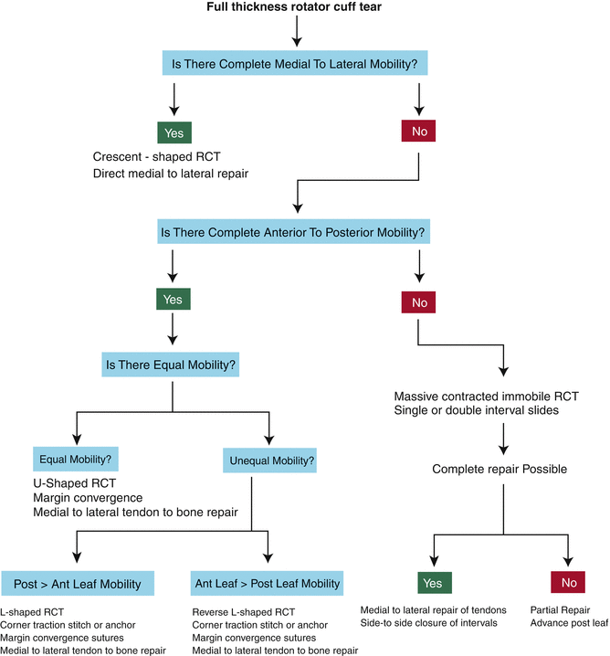
Table 25.1
The geometric classification of full-thickness, posterosuperior rotator cuff tears
Type | Description | Preoperative MRI | Intraoperative mobility | Treatment strategies |
|---|---|---|---|---|
1 | Crescent | Short and wide | Primarily medial-to-lateral mobility | End-to-bone repair |
2 | Longitudinal (L, rev-L, U) | Long and narrow | Primarily anterior-to-posterior mobility | Margin convergence, margin-to-bone repair |
3 | Massive contracted | Long and wide (>2 × 2 cm) | Relatively immobile in any direction | Interval slides, partial repair, load-sharing rip stop |
4 | Cuff tear arthropathy | GH arthrosis, proximal humeral migration | Completely immobile | Arthroplasty |

Fig. 25.3
Geometric classification system with treatment considerations. Intraoperative assessment of morphology and mobility of full-thickness rotator cuff tears allow the surgeon to make treatment decisions
Type 1 (crescent) tears have smaller length (ML dimension) than width (AP dimension); however, width varies greatly in size from small to massive. Crescent tears also have medial to lateral mobility that is sufficient for repair of the tendon directly lateral onto the bone bed under minimal tension (Fig. 25.4). These tears typically do not have significant medial retraction. Lastly, medial to lateral mobility should be equal along the length of the tear margin (Video 25.4).
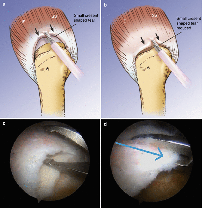

Fig. 25.4
Right shoulder. Crescent tear illustrations (a, b) and photographs (c, d) demonstrating the characteristics of width (AP dimension) greater than length (ML dimension), minimal retraction, and direct lateral mobility (blue arrow) of the tendon edge. SS supraspinatus, IS infraspinatus
Type 2 (longitudinal) tears have larger length (ML dimension) than width (AP dimension), and this width is typically <2 cm. The sub-classification of longitudinal tears as L-, reverse L-, and U-shaped is based on intraoperative assessment of the mobility of each leaf of the tear. L-shaped tears have a “corner” that is located along the anterolateral aspect of the posterior leaf of the tear (Fig. 25.5), and this corner will often have a “surprising” amount of posterior to anterior mobility that allows reduction to the anterolateral aspect of the bone bed (Video 25.5). In contrast, reverse L-shaped tears have a “corner” that is located along the posterolateral aspect of the anterior leaf (Fig. 25.6) and requires reduction in a posterolateral direction (Video 25.6). We have found that the corner of a reverse-L tear does not typically have the dramatic amount of anterior to posterior mobility that can be seen with L tears (opposite direction of reduction). In contrast, U-shaped tears have roughly equal mobility of the anterior and posterior tear margins without a clear “corner” to reduce (Fig. 25.7) (Videos 25.7 and 25.8). Recognizing longitudinal tear variations allows the surgeon to perform tension-free repairs using margin convergence sutures and/or suture anchors with margin-to-bone stitch configurations.
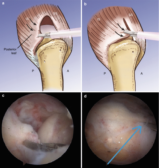
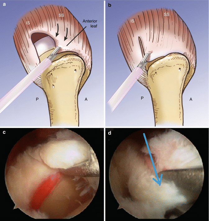
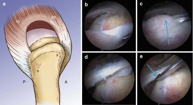

Fig. 25.5
Right shoulder. L-shaped longitudinal tear illustrations (a, b) and photographs (c, d) demonstrating characteristics of length (ML dimension) greater than width (AP dimension) and a “corner” that can be reduced to the anterolateral aspect of the bone bed (blue arrow). P posterior, A anterior, SS supraspinatus, IS infraspinatus

Fig. 25.6
Right shoulder. Reverse L-shaped longitudinal tear illustrations (a, b) and photographs (c, d) demonstrating characteristics of length greater than width and a “corner” that can be reduced to the posterolateral aspect of the bone bed (blue arrow). P posterior, A anterior, SS supraspinatus, IS infraspinatus

Fig. 25.7
Right shoulder. U-shaped longitudinal tear illustration (a). Photographs b, c show the direction of anterior mobility of the posterior margin, and photos d, e show the direction of posterior mobility of the anterior margin. P sterior, A anterior, SS supraspinatus, IS infraspinatus
Type 3 (massive contracted) tears are both long and wide, typically >2 cm × 2 cm (Fig. 25.8). These tears require advanced mobilization techniques for repair, because tendon mobility is poor in all directions (Video 25.9) [12]. Usually only partial repairs or single row repairs are possible for these tears. When assessing tear mobility with a tendon grasper, scarring and fibrosis may be based anteriorly, posteriorly, or in both locations. The anterior interval slide in continuity may be used to release adhesions that tether the cuff to the base of the coracoid via the superior glenohumeral ligament [13]. The posterior interval slide may be used to address posterior retraction and fibrosis [12]. Fibrosis of the joint capsule in a retracted tear may also be a cause of tear immobility; however, we have found that only a few millimeters of added lateral excursion is gained by a capsular release. In contrast, interval slides may result in several centimeters of added excursion.
