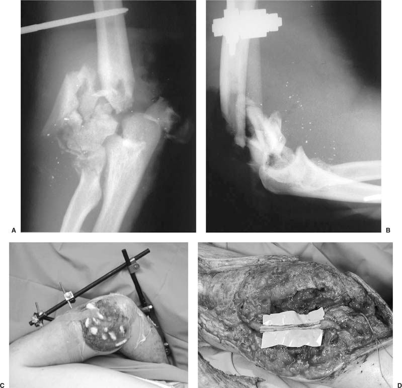
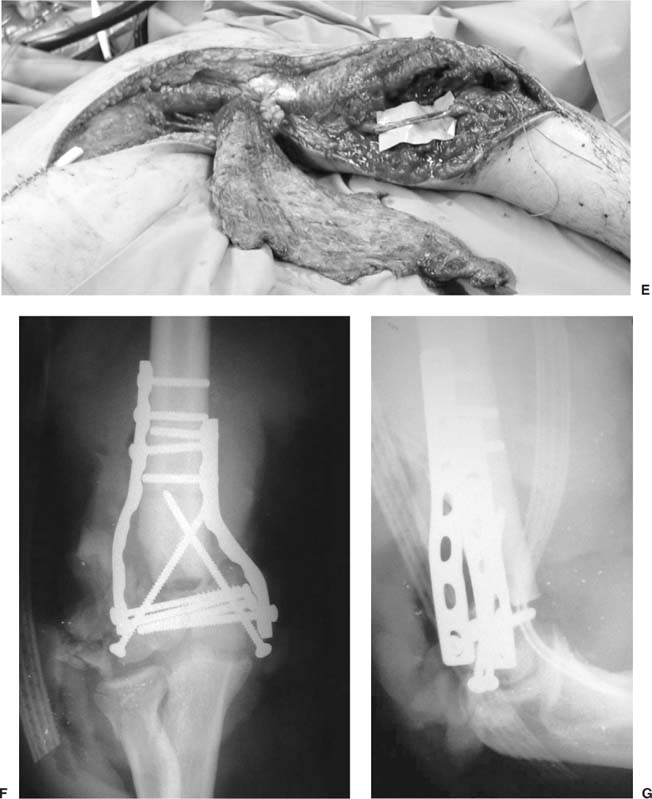
6 Open fractures about the shoulder and elbow are fairly uncommon. The consequences of open fracture that are most concerning are the increased risk of infection from bacterial seeding of the open wound. Damage to the surrounding soft tissue, and hence the vascularity and potential for union of the fracture, can play a role in open fractures about the elbow. The shoulder has a larger soft tissue envelope and is not as prone to open fractures. The principles of treatment in the shoulder and elbow are similar to open fracture treatment in other parts of the body. The goal is to achieve union of fractures without infection and restore normal function of the shoulder and elbow joint. Olson et al1 have outlined the course of treatment in patients with open fractures. Once the patient’s emergent and more pressing issues have been addressed, the initial treatment of the open fracture is the immobilization of the injured extremity and application of a sterile dressing to the wound. Intravenous antibiotics should be administered early, and the patient’s tetanus status should be updated. Urgent irrigation and debridement (I&D)with skeletal stabilization is then carried out and repeated, as needed, to achieve a clean bed of tissue. The wound is closed secondarily or covered with a flap when the patient is stabilized and the wound is adequately cleaned. These general principles are useful in the case of open elbow and shoulder fractures as well. Once the patient is stabilized, the wound dressed, and the extremity splinted, appropriate antibiotics should be started. Current recommendations are based on a modified Gustilo-Anderson2 open fracture classification system. Patzakis et al3 helped to establish the antibiotics that are used today. Most authors use a first-generation cephalosporin in types I and II fractures and a first-generation cephalosporin and an aminoglycoside in type III fractures. Penicillin is added for coverage of anaerobes in select, contaminated wounds (farmyard injuries). Most authors would recommend a course of 48 to 72 hours after I&D, and after each debridement and wound closure. The duration of antibiotic treatment, however, is controversial, and the protocols vary by institution. Routine cultures of open fractures at the time of initial I&D are probably not useful, as they do not typically correlate with the occurrence of infection or with the organism of infection.4,5 Cultures are typically used to identify organisms for specific treatment in draining or infected wounds. Debridement should be thorough and wide. The entire zone of injury should be visualized, and all devitalized and devascularized tissues should be removed. Exceptions are articular fragments, which should be cleaned, copiously irrigated, and saved for articular reconstruction. It has been suggested that in low-energy fractures, if the level of contamination is low (type I fracture) and there is a major piece of devitalized cortical bone that is essential to create an internal fixation construct, it may be retained after a thorough I&D.6 However, Edwards et al7 demonstrated that with retention of devitalized cortical fragments, infection rates increased by 50% in open tibia fractures. Once I&D is performed, the decision to perform definitive versus staged fixation should be made. Olson et al1 outlined the functions and goals of skeletal stabilization in open fractures. The length and alignment of long bones should be restored as well as articular surfaces displaced by fractures. Early use of the limb should be encouraged. Fracture union should be facilitated, and return of function should be allowed as soon as possible. These goals can be obtained with both internal and external fixation, and appropriate use of both of these techniques is paramount. In the case of intra-articular fractures (i.e., radial head and intracondylar humerus fractures), Mitchell and Shepard8 have shown that interfragmentary compression may greatly aid the healing of cartilage. Hence, the articulation may benefit from early internal fixation. The metaphyseal-diaphyseal junction may be spanned with temporary external fixation, and staged open reduction and internal fixation can then be performed. Articulated external fixators may allow early motion without internal fixation or with limited internal fixation. With grossly contaminated wounds, it is probably best to perform repeat debridements until the surgeon is confident of the wound prior to any sort of internal fixation. Fixation techniques for specific fractures are delineated in other chapters of this text and will not be dealt with further. Open fracture wounds should, in general, never be completely closed. There are some data on primary closure of open fractures, but there is always the risk of gas gangrene and clostridial infection with primary closure of open fractures.9 With type I open fractures, open wound care will generally lead to rapid secondary healing because the wounds are so small. Types II and IIIA wounds may require delayed primary closure or other procedures. Generally, primary closure at the time of I&D and fixation is not recommended for types II and IIIA wounds. Type IIIB wounds require flap closure by definition, and this is gone into greater detail elsewhere in this text. Antibiotic bead pouches are a useful temporizing measure in open fracture care. Various combinations of antibiotics and polymethylmethacrylate (PMMA) cement have been described. Ostermann et al10 described their use with open tibia fractures and decreased the infection rate of type IIIB fractures in particular (39 to 7.3%). The V.A.C. (Vacuum Assisted Closure) (Kinetic Concepts, San Antonio, TX) device has also begun to show promise as an adjunctive aid in the closure of open wounds.11 Biobrane (Bertek Pharmaceuticals, Morgantown, WV) and other occlusive dressings are also temporizing options for wound coverage. Infection in elbow and shoulder trauma is typically associated with attempts at open reduction and internal fixation and open fractures. They should be initially separated conceptually into acute and late infections. This is somewhat arbitrarily determined to be at the 3- to 4-week point. It is also important to determine if the fracture is healed or not. A late, healed infection may not require much more treatment than hardware removal, debridement, and appropriate antibiotic treatment. In the face of a chronically infected nonunion, however, the treatment course may become much more complicated. Patzakis12 describes four principles in the treatment of musculoskeletal infections, which may be useful when considering infection about the shoulder and elbow. The most important step in the eradication of infection is the debridement. All infected and nonviable tissues must be removed from the wound. Bone should be cut back to bleeding surfaces. Soft tissues should be debrided as needed to remove infected nonviable material from the wound. Attaining stable fixation construct to reduce motion and stabilize the wound is the next important step. Wound coverage should be achieved by local or free myocutaneous flaps to cover exposed bone or exposed joint surfaces and to increase vascularity. Healing of the fracture is the ultimate goal and can be aided with bone graft and other techniques as needed to aid in bony reconstruction and vascularity. Acute infection after open reduction and internal fixation of open fracture requires a high index of suspicion and vigilance. The wound that continues to drain should not be treated with “benign neglect.” Elbow and shoulder wounds typically do not drain for more than a few days to a week at most. Early motion is part of most treatment protocols and is not typically a cause of drainage. Hematoma and early infection are sources of persistent wound drainage in the shoulder and elbow. Literature from elbow arthroplasty series indicate that drainage after 10 days is significantly associated with infection.13 If caught early, irrigation and debridement with retention of hardware have a high chance of success. If the hardware is still providing stability and the fracture is not united, it is generally preferable to attempt to retain the hardware, although there is a risk of continued infection from organisms associated with the hardware. Motion of the fracture fragments is deleterious to the local vascularity of the fracture and soft tissue envelope. Therefore, if the infected hardware is not providing adequate stability, it should be removed, and an alternate means of fixation (such as an external fixator) should be used. Closure of the soft tissues should be performed over drains. Appropriate cultures from multiple sites should be taken prior to the use of antibiotics in the irrigation.14 Broad-spectrum antibiotics should be initiated after cultures are obtained, then tailored to the culture results as soon as possible. Treatment duration varies, but we suggest 6 weeks of intravenous treatment for patients with established osteomyelitis. If the wound cannot be closed without tension, an antibiotic bead pouch or other occlusive dressings can be used for temporary coverage until a more definitive solution is obtained (i.e., myocutanous flap). Soft tissue coverage is typically more of a problem about the elbow than the shoulder. Unless there is an open injury with loss, the soft tissues about the shoulder tend to be fairly resilient, and skin slough is uncommon. About the elbow, however, skin coverage is more tenuous. The use of multiple incisions and the failure to maintain full thickness flaps may lead to necrosis. There is relatively little muscular coverage over the elbow articulation itself. With infection and loss of soft tissues, staged coverage may be necessary. Antibiotic bead pouches are useful to aid in the creation of a healthy recipient bed for flap coverage. Flap coverage techniques are covered in Chapter 7 for the elbow and Chapter 15 for the shoulder. In the face of chronic infection, hardware removal is mandatory. If the fracture is healed, then this is not a problem. However, when there is a chronically infected nonunion, the scenario is somewhat more complicated. Typically, the hardware is no longer serving its purpose. Therefore, it is only serving as a nidus to perpetuate the infection. Proper irrigation and debridement will mandate removal of the hardware. Any devitalized tissues should also be removed. After a thorough debridement, the next stages are dependent on the presence or absence of osseous union. If the fracture is healed, then wound coverage and antibiotic course may be addressed. If there is a nonunion, stabilization may require temporary spanning external fixation. This allows for stabilization of the joint to allow for soft tissue rescue and infection control. The articulations themselves must then be evaluated. If there is considerable damage to the articular surfaces themselves, consideration must be given to what type of ultimate outcome will best benefit the patient. Arthrodesis is an option for both the shoulder and the elbow when the articular surfaces are in poor condition or the fracture is not considered reconstructable. It is typically considered in patients who have had previous chronic infections, but have otherwise functional limbs. Fascial arthroplasty with distraction fixation is a consideration in a patient if there is a need for a mobile articulation and the infection is eradicated. Most experience with this is in the young high-demand patient with arthritis of the elbow.15 If the articular surfaces are in good condition, after infection control is obtained, the next stage is to move on to bony reconstruction. For supracondylar fractures of the distal humerus, bony shortening and bicondylar fixation can give good results and obviate the need for structural bone grafts or bone transport techniques. The articular surface should first be reconstructed using lag screws if there is no comminution. In the presence of articular comminution, the screw fixation of the joint should not be in lag fashion. Humeral shortening of up to an inch is well tolerated and is a useful technique to overcome metaphyseal comminution and bone loss.16 Bicondylar fixation is a proven technique. We prefer fixation on the direct medial and lateral surfaces, whereas others have supported fixation of the medial and posterolateral surfaces.17,18 With severely osteopenic bone, locked plating and bone augmentation with PMMA or Cortoss (Orthovita, Malvern, PA) may be useful. Ilizarov techniques have also been described and may be useful for overcoming osteopenia and bone loss.19 In the case of infected radial head and olecranon fractures, resection is a viable alternative to attempted reconstruction. Infection in association with radial head open reduction and internal fixation or replacement, however, seems to be uncommon. A recent series of radial head replacements for trauma had 1 infection (superficial) out of 25, and it was salvaged without implant removal.20 In radial head injuries, the integrity of the interosseous membrane must be established. If the interosseous membrane is intact, then resection can be safely undertaken without worry of radial migration and late wrist pain. Goldberg et al21 reported satisfactory function in over 90% of patients with isolated radial head injuries treated with excision. If the interosseous membrane is disrupted, then consideration to adjunctive procedures to address the issue of radial migration must be undertaken. Edwards and Jupiter22 reported on a small series of patients that had Essex-Lopresti lesions and radial head fracture. They treated unsalvageable radial heads with Silastic replacement and ulnar shortening with good results. Because Silastic implants have been shown to not be able to withstand physiological loads, ulnar shortening was probably necessary in some of the patients to reestablish distal radioulnar joint kinematics. With modern metallic implants, it is thought that there is less chance for subsidence. This would obviate the need for ulnar shortening procedures to maintain the relationship of the radius to the ulna. Radial head replacement can be considered in a patient who needs restoration of radial length and a clean wound bed. The infected olecranon nonunion is probably best treated with excision and triceps advancement. Olecranon excision is a well-described procedure for treatment of even acute olecranon fractures. An et al23 described the limits of olecranon excision with respect to elbow stability/constraint. In the case of an infected olecranon nonunion, as in acute fractures, upwards of 50% can usually be excised with a triceps advancement and maintain elbow stability. Some authors have suggested that up to 80% can be excised, but An et al demonstrated a linear decrease in elbow constraint with increasing fragment size−50% seeming to be the point at which instability would result. Gartsman et al24 found there to be no differences in isometrics between patients treated with excision versus open reduction and internal fixation; it appears that excision is a viable, functional alternative to staged open reduction in the face of infected nonunion of the olecranon. With fragments that exceed this size (50%), treatment may need to be individualized. If the elbow is unstable, staged fixation may be necessary with use of hinged fixators to maintain stability as needed. Infected nonunions about the shoulder may be problematic. Greater tuberosity nonunions, once sterilized, may be treated with fixation and bone grafting as necessary. Direct repair of the supraspinatus tendon to the humerus may be necessary if the tuberosity is not salvageable. More complex fractures may or may not be reconstructable and would lead to a recommendation of arthrodesis. We would caution against the use of hemi or total shoulder arthroplasty to salvage the shoulder articulation in the face of previous infection. As in other parts of the body, a history of previous infection makes arthroplasty a risky reconstructive option, though not impossible. Complete resolution of the infection is necessary. In select low-demand patients, Girdlestone-type procedures may work best. Open fractures, comminution, high-energy injury, infection, bony defects, and poor fixation are some of the factors that predispose shoulder and elbow fractures to poor results.25–28 Fractures about the shoulder and elbow can be challenging, and poor attempts at primary fixation can lead to difficult reconstructive problems. Bony defects from open injuries, severe comminution, and/or infection can add to the difficulty of treating these fractures acutely. Treatment options in both the shoulder and the elbow are repeat open reduction and internal fixation with or without bone graft, arthroplasty, arthrodesis, or allograft replacement. In the elbow, hinge fixators and Ilizarov techniques may be useful to augment stability and fixation or to help overcome bony deficits. Supercondylar/intercondylar mal/nonunions can often be treated with repeat open reduction and internal fixation. Humeral shortening of up to 3 cm is a useful technique to obtain healthy bone edges and easier surfaces to compress. We recommend rigid fixation of both columns (lateral and medial) with well-contoured plates using balanced AO method fixation. In severely osteopenic bone, locking plate constructs are useful because they are not as dependent on bone quality for fixation. Bone grafts are mandatory and may include impaction grafting of the humeral canal to avoid placement of graft in articular areas. In the case of severe bone loss of a single column of the supracondylar humerus, cortical grafts may be useful. There are several precontoured distal humeral plates now available that may provide a ready template to reconstruct the distal humerus. McKee et al29 reported on a series of 13 patients who had mal-and nonunions of intra-articular fractures of the distal humerus. These patients all had significant pain and dysfunctional elbows. McKee et al’s approach was to first carefully assess the residual deformity and arthrosis of the elbow. Osteotomy was performed for malunion. Debridement was performed for nonunion. Stable fixation with bone grafting as needed was then performed. Anterior and posterior releases, with ulnar nerve transposition/neurolysis, were then performed as indicated. McKee et al’s series reported a doubled range of motion (to 90 degrees) and 10 of 13 excellent or good results using the Morrey elbow score. The elbow is often stiff on presentation and may require an extensive release to regain motion. A hinged fixator may be necessary after this release to stabilize the elbow and to allow early motion. A successful bony reconstruction can still end up with a stiff nonfunctional elbow if early motion in an adequate arc is not achieved. If the patient has a significantly frozen elbow prior to bony reconstruction, it is wise to anticipate the need to perform a soft tissue release. This may in fact destabilize the elbow, which will require hinged fixator placement to allow for early motion. This has been recommended for use in selected cases, although there is no reported case series to our knowledge. There are reports of using the hinged fixator successfully with difficult elbow instability problems.30 In general, an extensile posterior approach is best for these problem fractures. If there is no intra-articular involvement, the intervals on both the medial and lateral sides of the triceps can be developed to expose the humerus. The ulnar nerve should be transposed to allow for better access to the medial column and elbow joint as necessary. The radial nerve should be identified and protected. Greater tuberosity mal-and nonunions can be problematic. Small amounts of displacement may cause impingement of the tuberosity in the subacromial space. Craig31 recommends treatment when there is either significant displacement of the tuberosity or a functional deficit secondary to pain or limited range of motion. The fragment is generally displaced superior or posterior or both. CT is often very useful to determine the location of the fragment relative to the anatomic origin of the fragment. It is commonly difficult to assess posterior displacement on plain radiographs. Superior displacement is more readily evident on plain radiographs. Displaced and nonunited greater tuberosity fragments shorten the rotator cuff and capsule over time. These structures will require extensive mobilization to anatomically reduce the fracture with chronic presentation. This will require more exposure than is typically necessary to fix an acute fracture. Some authors31 recommend using a deltoid-splitting approach extended by taking down the anterior deltoid. We have used a deltopectoral approach with success. Extra and intra-articular adhesions will need to be taken down. The greater tuberosity fragment should be handled with care, although it will need to be osteotomized in the case of malunion. Once the fragment is adequately mobilized, the rotator interval may be closed to help reapproximate the fragment. It is useful to place several heavy sutures in the bone-tendon junction to better control the fragment. The recipient bed should be decorticated to restore a bleeding bed of bone. Typically, the recipient bed is sclerotic in the chronic mal-and nonunion. Iliac crest bone graft should be harvested as needed, particularly in the case of nonunion. The greater tuberosity fragment may need to be contoured to better fit the recipient bed. Fixation methods vary from tension bands to screw fixation using AO methods. Method of fixation is typically dependent on the quality and amount of bone present. The goals of treatment in both the shoulder and the elbow are to restore motion/function, heal the fracture, and reduce pain. These considerations should remain paramount at all times. Therefore, any procedure that is contemplated should attempt to address all of these issues simultaneously. Principles of treatment include release of contractures (about the elbow), correction of deformity, stable fixation, and stimulation of healing with bone grafting. When stable fixation is unlikely, or when the patient is low demand/elderly, an arthroplasty may be considered. In higher demand or younger patients with loss of the elbow articulation, fascial arthroplasty may be considered.15 Shoulder or elbow fusion is controversial but should be considered in select patients with soft tissue problems and infections in particular. A recent study by Tang et al32 suggests that 110 degrees of flexion is optimal when fusing one elbow with nonrestricted shoulder, wrist, and hand motion. Extra-articular gunshot wound fractures about the elbow and the shoulder may not need treatment beyond appropriate antibiotics, as well as bracing as indicated with early motion. Indications for fixation are similar to those for closed injuries. Sarmiento et al33 described the closed treatment of extra-articular distal humerus fractures. This has been successful in the authors’ hands as well. In Sarmiento et al’s33 series, there were 8 gunshot wound fractures out of 85 distal humeral shaft fractures treated with functional bracing. There were no infections or mal/nonunions per their criteria. Fracture bracing was initiated on average on day 12. Gunshot wounds of the articulations of the shoulder and elbow require careful evaluation to rule in or out the presence of bullet fragments in the articulation itself. Traditionally, open arthrotomy was performed to remove these fragments, but arthroscopic techniques now allow for a minimally traumatic method of debriding the joint and removing foreign bodies. Certainly lead resorption by the synovial fluid has been well documented, as well as the detrimental effects of third-body wear in the articulations themselves.34 Jamdar et al35 reported arthroscopic removal of shotgun pellets from the elbow. Arthroscopic treatment of injuries about the shoulder is expanding. One interesting injury of the shoulder girdle is the so-called floating shoulder or double disruption of the superior shoulder suspensory complex. Traditionally, this has been viewed as an injury that mandates surgical treatment.36 However, recent series have called this into question. Edwards et al37 found good results with nonoperative treatment of two patients who sustained floating shoulders from gunshot wounds. One patient had a clavicular nonunion with segmental bone loss but still had a satisfactory result. Egol et al38 could not elicit a significant functional benefit from operative treatment of floating shoulder injuries as well. Currently, we would caution against nonoperative treatment if there were segmental bone loss. As Edwards et al37 recommend, however, if the patient is able to cooperate with an early motion program, nonoperative treatment may be entertained. Antibiotic treatment of gunshot wound fractures varies from institution to institution. Knapp et al39 established the following guidelines that we find useful. For patients who are not otherwise requiring surgical stabilization, a 3-day course of a fluoroquinolone and local wound care are adequate treatment. If the patient’s fracture requires surgical stabilization, a 3-day course of a first-generation cephalosporin and an aminoglycoside are given. These guidelines are for low-velocity gunshot wounds only. High-velocity gunshot wound injuries will invariably require operative debridement and further treatment of fractures as indicated. When supracondylar humerus gunshot wound fractures are encountered, one of the major concerns is the vascularity of the extremity. Brannon et al40 recommended angiography for all of these injuries, although in their series all 13 patients had normal angiography. Despite their series of negative angiography, concerns about simple observation remain. Drapnas et al41 found a 27% incidence of palpable distal pulses and positive major arterial injury in their series. Snyder et al42 found that angiography had a 92% accuracy in diagnosing arterial injury and no significant morbidity in the penetrating trauma patient population. Given these data, in a patient with an obvious arterial injury, urgent surgical exploration, temporary skeletal stabilization, and revascularization are recommended, in that sequence. In the patient with a patently normal vascular exam and normal side-to-side brachial indices, observation is prudent. However, the patient with diminished pulses and mild signs of ischemia should undergo angiography. Fasciotomy of the distal extremity is recommended in general for patients who require a vascular repair/revascularization. Compartment syndrome is certainly a risk for a patient with prolonged forearm ischemia, and the sequelae of an untreated compartment syndrome may be devastating. Secondary closure versus skin grafting may be performed on a delayed basis.
Open Fractures, Infections, Non/Malunion, and Heterotopic Ossification of the Shoulder and Elbow
Open Fractures
Infection
Nonunions/Malunion
Fractures Resulting from Gunshot Wounds
< div class='tao-gold-member'>
Open Fractures, Infections, Non/Malunion and Heterotopic Ossification of the Shoulder and Elbow
Only gold members can continue reading. Log In or Register to continue

Full access? Get Clinical Tree


