Open fractures
Specific open fracture types in the elderly
INTRODUCTION
Our population is aging with the number of people in the United States older than 65 expected to double to 60 million or 17% of the population by 2030.1 Compared to the past, this is a more active population with an increased lifespan and has been working and maintaining their independence for longer. In addition, in 2010 the United States census found that the population aged between 45 and 64 had a growth rate of 32%, primarily due to the baby boomers.2 This population growth will be reflected in the years to come in North America.
The increasingly elderly population can be expected to lead to an increased number of trauma patients over 65. Trauma is currently the fifth leading cause of death in those above 65 and accounts for 23% of hospital admissions and 28% of all hospital charges.1 This will have significant implications for future healthcare cost as the population grows. The elderly generally have more medical comorbidities, and so longer lengths of stay can be anticipated after trauma.
The geriatric population has been well studied in terms of certain types of fractures including distal radius and hip fractures. However, geriatric open fractures are not a well-researched subset of injuries. In the past, it has been hard to record large enough numbers to evaluate these injuries although the current literature is increasing studying geriatric traumatic fractures. With the growing elderly population, these fractures will affect many healthcare disciplines, including the subspecialty of orthopaedic trauma.
EPIDEMIOLOGY
Despite the aging population and frequency of fractures among the elderly, the literature on open fractures in this group of patients is limited. We do know that unlike open fractures in the young, 60% of open fractures in the elderly occur due to low energy mechanisms such as a fall. The most common open fractures are sustained in the distal radius, ankle, fingers and the tibial diaphysis. Much of the information we have on epidemiology comes from the Royal Infirmary of Edinburgh and the work of Court-Brown et al.3 Prospective data on fractures occurring in 4,786 consecutive outpatients and inpatients ≥65 years of age were collected over a 2-year period. The majority of fractures in the elderly population were in women (77%), and only 1.2% were open fractures, 13.6% of which were Gustilo type III. This trend towards a higher incidence of Gustilo type III open fractures would typically be expected as the energy mechanism increases, although in the elderly they can occur after ground level falls.
Court-Brown et al.4 reviewed 484 open fractures in patients ≥65 years of age over a 15-year period and compared them to open fractures in patients below 65. The incidence of open fractures was noted to increase with age. Interestingly, males aged 15–19 and females ≥90 showed a similar incidence of open fractures. These results are from the United Kingdom and may be different elsewhere. However, females were noted to have an increase in open fractures throughout their life, but with a sharp increase starting in the seventh decade. This could be due to a physical decline with decreased bone mineral density, and decreased physical activity leading to balance issues perpetuating a vicious cycle. The main mechanism of injury was a fall in 60% of patients, representing a low energy mechanism, yet there was a high incidence of type III fractures. This was true especially in women. There is a definitive increase in open fractures in post-menopausal women. The decline in bone quality in post-menopausal women is also accompanied by changes in the skin.
Animal studies show that the male dermis is 190% thicker than in the female, while the epidermis is thicker in female mice.5 This is similar to the situation in humans.6 Applying this to the results of a simple ground level fall, the higher grade of open fracture can possibly be attributed to the skin quality in elderly patients and may not be reflective of the energy of the injury. The skin is thinner and may tear more easily, resulting in a higher grade of open fracture due to the size of the wound (Figure 13.1).
Cox et al.7 specifically compared the comorbidities and mechanism of injury for tibial shaft fractures sustained in the elderly. Comorbidities were found to be similar between patients sustaining closed or open fractures. The mechanism of injury was also similar, with ground level falls and pedestrian versus motor vehicle accidents predominating. Patients with Gustilo type III open fractures had more complications, and increased length of stay and need for additional surgery compared to those with lower Gustilo types.
Keller et al.1 retrospectively reviewed all high energy trauma in patients ≥65 over a 5.5-year period at their academic trauma centre. Clavicle, midfoot, proximal humerus, sacroiliac joint and distal ulna fractures were predictors of mortality in geriatric patients. It was also highlighted that the elderly present with worse injuries, remain in hospital longer, require greater resources after discharge and die at a rate three times higher than the younger population after high energy trauma. The impact or frequency of open fractures in this study population of 597 was not studied.
In a similar study, Abdelfattah et al. reviewed 154 geriatric patients, admitted over a 6-year period, who sustained high energy trauma (Injury Severity Score [ISS] >16).2 Clavicle, scapula and femur fractures were found to predispose this population to worse outcomes. Increased mortality was found in those with pelvic or acetabular fractures requiring operative fixation and spine fractures that were treated non-operatively. Again no assessment of those patients with open fractures was delineated.
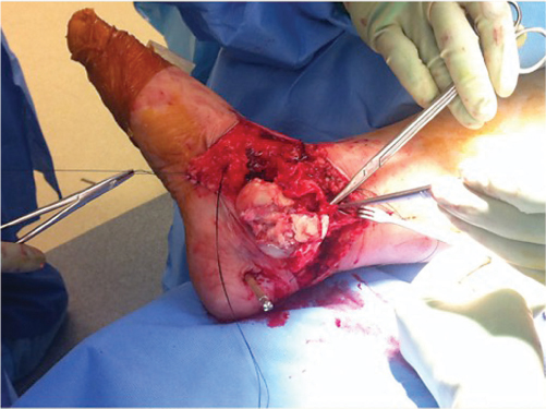
Figure 13.1 Geriatric open fracture with large open wound due to skin tearing.
In this chapter we hope to draw attention to the important issue of geriatric open fractures as there is very little literature to guide care. Several articles do discuss geriatric trauma, both low and high energy, but open fractures are frequently not mentioned or evaluated.
CLASSIFICATION
The Gustilo classification of open fractures has been commonly utilized to grade all open fractures. In this system, a type I is an open fracture with a clean wound less than 1 cm long, a type II has a laceration over 1 cm long without extensive soft tissue damage and type III fractures have more extensive soft tissue damage. Type III fractures are subdivided into IIIA, IIIB and IIIC fractures. Type IIIA fractures are those which can be closed primarily. Type IIIB fractures require the soft tissue injury to be closed secondarily by skin grafting, flaps or other techniques. Type IIIC fractures are those with a vascular injury requiring repair.8
The Gustilo classification has been used extensively. However, in the elderly population even the mechanism leading to the open fracture is not the same as in younger patients. Often the geriatric patient has very thin skin, which may contribute to larger wounds. As one ages, there is an increase in soft tissue fragility. In addition to the changes which we know occur with aging of the elderly bone and the decrease in bone mineral density, elderly skin has decreased organization of collagen making it more susceptible to injury.4 The skin loses elasticity and often feels ‘loose’ without significant connection to the underlying soft tissue. In addition, the skin turgor is not the same as in the younger patient. Thus, the older patient may have propagation of skin trauma from a small bony insult yielding a much larger skin tear. As the skin tears, it causes a much higher grade injury due to the larger size of the soft tissue injury.
A common example is an open ankle fracture dislocation. An eversion type of mechanism is common. The medial malleolus fracture may be quite small but the wound presents as a transverse laceration 10 cm or more in length. This is because the skin is quite fragile and tears more easily. The open fracture classification of Gustilo and Anderson is useful in the younger and high energy trauma patient, but in the geriatric fracture patient, its applicability may be more limited. Surgeons should perhaps consider an alternative classification which evaluates multiple factors.
The value of classification systems for mangled extremities was evaluated in the Lower Extremity Assessment Project (LEAP) study.9 There are several classifications for mangled extremities taking into consideration the injury to the skin, muscle and nerves; ischaemia; chronological age; energy of injury; and other factors. Those scoring systems were used to predict amputation versus limb salvage. The various scoring systems proved unreliable in predicting outcome.10 In the elderly open fracture we have already established that there often is a higher grade of open fracture due to the size of the wound. The literature is limited but suggests that outcomes in higher fracture grades are worse.7
Table 13.1 Geriatric open fracture considerations to further assess severity of the fracture

Table 13.1 shows a number of additional factors which, we believe, should be taken into consideration when classifying elderly open fractures. These are all questions relating to the injury, which should be answered to assist in planning the treatment of the elderly open fracture. We suggest evaluation of these additional factors when considering the severity of open fractures in the geriatric population.
TREATMENT
As with any open fracture there are several considerations before operative fixation. The first involves a thorough clinical evaluation consisting of the basic principles of Airway-Breathing-Circulation (ABC) management. Early resuscitation is essential for decreasing mortality in this often fragile population. Once the patient is stabilized, exposure of all injured extremities will allow complete assessment of the viability of the limb, degree of soft tissue damage and amount of contamination present. Compartment syndrome cannot be excluded despite the open nature of the fracture or because of the age of the patient. If vascular injury is suspected, ankle brachial indices (ABIs) should be used as a screening tool. Complete radiographic evaluation of the fractured limb to include the adjacent joints is essential. In periarticular fractures, CT scans can be very useful after spanning external fixation has been applied as they may reveal fracture morphology better than X-rays. This can be indispensable for preoperative planning.
Once the general assessment is complete, specific emergency room management should be tailored to the fracture and soft tissue injury. Wound haemorrhage must be controlled. If the bleeding is from one of the extremities, direct pressure is preferable with application of a tourniquet if the former is insufficient. If there is an open pelvic fracture, intra-pelvic packing may be necessary. If the pelvic fracture is an open book fracture, a binder or sheet may be required, applied over the greater trochanters to decrease blood loss.
Antibiotic administration is also important in the emergency room. The antibiotics will depend on the initial classification of the open fracture. This has been historically based on the Gustilo and Anderson classification (Table 13.2). However, in the elderly, with an increased risk of renal dysfunction and potential for decreased creatinine clearance, the choice and dose of antibiotic regimens may need to be altered.
Table 13.2 Antibiotics recommended for different fracture grades as defined by the Gustilo–Anderson Classification
Open fracture type | Antibiotic recommendations |
|---|---|
Type I | First generation cephalosporin |
Type II | First generation cephalosporin |
Type III | First generation cephalosporin + aminoglycoside + penicillin for farm injuries |
Fractures will then need provisional irrigation and reduction followed by placement in a properly padded splint before transfer to the operating room. Formal irrigation and debridement (I&D) in the operating room should be initiated as soon as deemed safe by all care teams, and haemodynamic stability achieved. Meticulous assessment of the zone of injury is important, with a systematic debridement being undertaken. This should start with the most superficial structures and proceed down to the bone. Any non-viable structures or foreign bodies should be excised. Extension of the wound is usually necessary for complete assessment.
Irrigation with normal saline should follow debridement. No advantage has been shown with the addition of antibiotic solution to normal saline irrigation. Debate continues as to whether there is an advantage to pulsatile lavage versus low flow irrigation. Some surgeons feel that tissue is traumatized with pulsatile lavage and foreign material is driven further into the wound than with gentle high volume irrigation. In the elderly, the skin is thinner and more friable making it less tolerant of even minimal tissue trauma. This favours low flow irrigation.
Based on the amount of contamination and extent of injury at initial I&D, a decision regarding wound closure or repeat debridement must be made. The level of soft tissue viability following a high energy mechanism of injury may not be apparent on initial debridement and a second look is likely to be beneficial. The fragility of the soft tissues and potential vascular compromise by pre-morbid conditions in the elderly may make even a low energy injury best managed with a second look. Other situations where this may apply is in a moderate to severely contaminated wound, when there has been a delay in transfer to the operating room due to polytrauma or comorbidities, or in a damage control situation where the time required for wound closure places the patient at greater risk. In these situations negative pressure wound therapy (NPWT) allows for control of drainage at the site of injury, provides a sterile environment and allows ease of exposure on return to the operating room (Figure 13.2). Closure may follow when the wound is felt to be clean and there is no concern about further progression. The use of vessel loops sequentially crossing the wound and secured with staples tends to ensure better approximation of the wound edges as opposed to the sponge alone which tends to push the skin edges further apart. The negative pressure device has adaptations that may protect fragile elderly tissue. The pressure can be lower than the typically used 125 mmHg especially if placed over nerves or vessels, or traumatized muscle tissue. Different sponge types or dressings beneath the sponge provide added protection. Intermittent suction has the same benefits of tissue protection as the continuous mode.
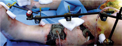
Figure 13.2 Use of negative pressure wound therapy for an open fracture.
Some debate exists in the literature as to whether post-irrigation wound cultures should be obtained. Post-irrigation wound cultures have been used to guide antibiotic therapy should a deep infection develop after treatment of an open fracture. These have proven to be more accurate than pre-irrigation cultures. Lenarz et al.11 retrospectively reviewed 346 open fractures at a single level 1 trauma centre. A standard protocol was utilized to determine timing of closure for open fractures. Type II and III fractures were included as the majority of type I fractures were closed primarily after initial I&D. Post-irrigation cultures were taken and an NPWT device applied. If the cultures remained negative at 48 hours, the wound underwent repeat I&D and closure. If the cultures were positive at this point in time, the wound underwent a further I&D and the NPWT was replaced. This process was repeated until the cultures remained negative, at which time the wound was closed. The authors found a decrease in deep infection across all open fracture grades when compared to historic controls.
If the injury is amenable to primary closure, many factors must be considered. As soft tissue in the elderly is prone to swelling, early meticulous closure can avoid future complications and decrease the need for plastic surgery coverage. The basic principles of suturing are even more important in the elderly. One must avoid strangulation and prevent necrosis of the tissue edges. Many aspects should be thoughtfully evaluated for the best outcomes. The use of a reverse cutting needle can prevent tearing of the suture through the skin towards the wound edge once tension is placed. Also, entering the skin at a 90 degree angle minimizes the tissue trauma of needle placement and allows proper eversion of the skin edges. The use of non-toothed rather than toothed forceps can prevent crushing the tissue as sutures are placed. Various suture techniques can also be used to aid closure. At times multiple closure techniques will need to be combined for the best closure (Figure 13.3). Use of far-near near-far retention sutures spaced along the incision is useful when wound approximation is difficult and can decrease the tension on any one suture, preventing them from tearing fragile skin. These can be left in for a period of time until the tension has decreased, or on initial closure once the remaining sutures are placed. Horizontal mattress sutures perform the same function of distributing skin and suture tension while everting the skin edges for improved healing (Figure 13.4).
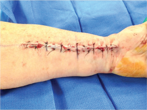
Figure 13.3 Multiple suture techniques used to close an open both-bones forearm fracture.
Related posts:

Stay updated, free articles. Join our Telegram channel

Full access? Get Clinical Tree


