Neurological System

11


Neurological System


Katie M. Walsh and Tamara C. Valovich McLeod


Introduction


Neurological disorders in athletes are generally placed in two categories: life-threatening conditions such as encephalitis, meningitis, or stroke; and those with chronic implications, which may include Guillain-Barré, multiple sclerosis, migraines, amyotrophic lateral sclerosis (ALS), complex regional pain syndrome (CRPS), or epilepsy. In addition, there has been increased emphasis on sport-related concussion because of the incidence of and possible short- and long-term consequences of concussive injuries. Because of the age range and close living quarters shared by many college athletes, they are more susceptible to certain neurological diseases, such as meningitis. In addition, outdoor practices in the late afternoon warm weather are favorable to mosquitoes, the carrier of certain types of encephalitis. The health care provider who appreciates these conditions will be better able to recognize and possibly prevent contact with and spread of these deadly disorders. Infectious neurological conditions (encephalitis and meningitis) are discussed in Chapter 15.


Although chronic neurological conditions are less common, they produce symptoms that can interfere with normal daily function as well as prevent vigorous activity for periods of time. Most have no clear or distinguishable signs, and only the patient’s particular symptoms can guide the clinician to a correct impression. These conditions can also cause emergent situations that require immediate medical referral.


Many symptoms associated with neurological conditions are vague and fleeting and are sometimes passed off as a result of overtraining or fatigue, both of which are common to the athlete. This chapter reviews the pertinent neurological anatomy and highlights signs and symptoms associated with specific neurological conditions. A strong knowledge base will enable the athletic trainer to recognize important symptoms and properly refer the athlete to a physician.


Neuroanatomy


Skull


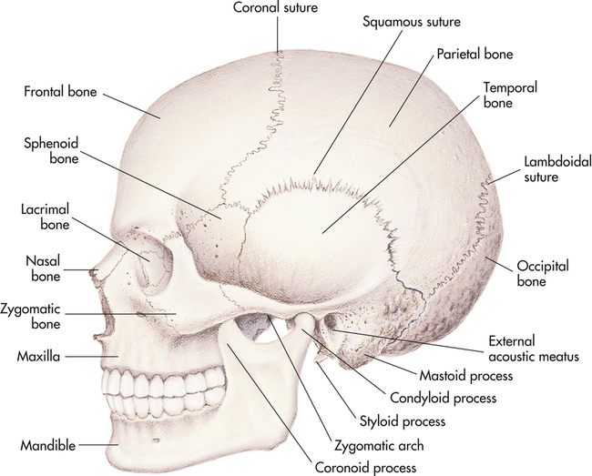
The human skull is made up of two main components: the cerebral cranium, which protects the brain and brainstem, and the anterior facial bony structure. The cerebral cranium consists of eight bones that include the frontal, two temporal, two parietal, occipital, sphenoid, and ethmoid bones. The facial skeleton is made up of the mandible, two zygomatic, two maxillary, and two nasal bones (Figure 11-1).


Meninges


The meninges lie just under the skull and provide three protective layers: the outermost dura mater, the arachnoid membrane, and the pia mater. The dura mater is a thick, tough, fibrous membrane functionally composed of two layers with the periosteum against the skull and the inner dura supporting structures of the brain. The real and potential spaces formed between these membranes allow for arteriovenous connections that can be disrupted by hematomas (i.e., accumulations of blood between the spaces) caused by trauma.


Blood supply to the meninges comes from vessels that follow grooves in the skull. Although the periosteum adheres directly to the skull bones, a potential epidural space exists and ruptured vessels or infection can cause an actual space to form as seen when a hemorrhage causes an epidural hematoma. In the spine a true epidural space, separating the dura from the periosteum of the vertebral bones, contains fat and epidural veins.


The inner lining of the dura mater forms folds in the cerebral hemispheres. The membranous plate known as the falx cerebri divides the hemispheres into right and left halves as seen in Figure 11-2. Another plate formed by the dura, the tentorium cerebelli, separates the cerebral hemispheres from the cerebellum and brainstem.


Large sinuses or cavities that lie within the dura mater allow for the venous return from cerebral veins located between the two layers. The walls of these sinuses are made up primarily of dura. The function of the superior sagittal sinus is to collect venous blood, as well as excess cerebrospinal fluid (CSF), which drains through its arachnoid villi. The blood flows into the transverse sinuses, which also receive blood from other veins of the brain. Together with the sigmoid sinuses, these major venous pathways leaving the brain become the internal jugular vein.


The cavernous sinuses are smaller and receive venous blood from the hypothalamus. They are of clinical importance because the internal carotid artery and several nerves pass through them after entering the cranium at the base of the skull.


The arachnoid membrane is named for its delicate, spiderweb-like consistency. The arachnoid membrane and dura mater are separated by the subdural space, which is a potential space that contains only a few drops of CSF and numerous small veins. If the membrane is disrupted, it can become distended with blood, pus, or other fluids during pathological conditions. The subarachnoid space separates the arachnoid from the pia mater and is filled with fibrous connecting trabeculae, the major arteries, and CSF. Because the trabecular connections are subtle, the arachnoid and pial layers are sometimes called the leptomeninges.


The pia mater is the innermost layer of the meninges and is very thin, adhering directly to the surface of the brain and spinal cord. It follows every fold and enters every crevice, making it difficult to distinguish this membrane from the surface to which it adheres. No potential or actual spaces exist between the pia mater and neural tissue.


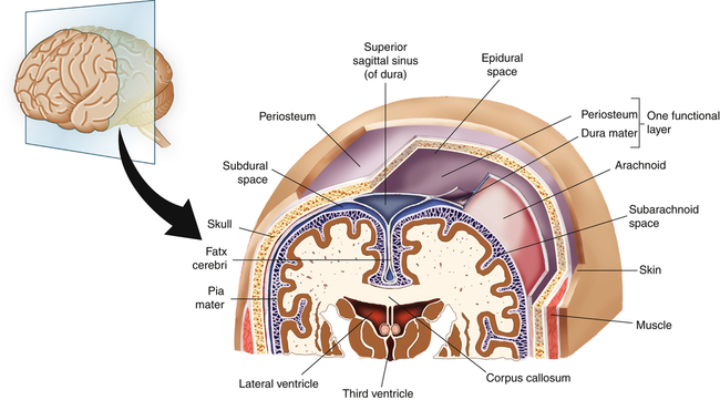
Meninges also provide a protective covering to the brainstem and spinal cord.


Cerebrum


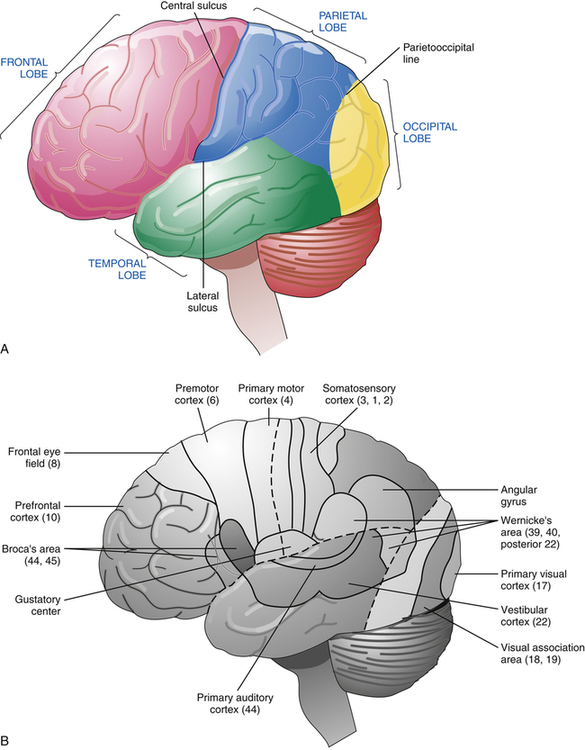
The two cerebral hemispheres are composed of neural tissue. These hemispheres are divided into four principal lobes: frontal, temporal, parietal, and occipital (Figure 11-3, A). The lobes have been extensively researched in attempts to isolate the locations of specific brain physiological functions and pathological processes. The most commonly used classification is Brodmann’s classification system, which identifies specific functional cortices of each lobe by numbers (see Figure 11-3, B).


The frontal lobe contains the primary motor area (area 4), the premotor area (area 6), the frontal eye field (area 8), Broca’s speech area (areas 44 and 45), and the frontal association area (areas 9, 10, and 11). The primary motor cortex is highly organized in a somatotopic fashion with the lips, tongue, face, and hands on the lowest part, moving upward to trunk, arm, and hips, and ultimately to the foot, lower legs, and genitalia that hang over the edge into the interhemispheric fissure. This cortical area is responsible for voluntary movement of skeletal muscle of the contralateral side of the body. The frontal eye fields coordinate contralateral deviation of the head and eyes.


Clinically, seizure activity within this cortex results in convulsions of the parts of the body represented in the area of electrical disruption. Damage to this cortex can lead to contralateral flaccid paresis or paralysis, and spasticity is usually present if there is concomitant injury to the premotor area. The left hemisphere usually influences and correlates with right hand dominance. Broca’s area is located in the dominant hemisphere only, and, when this area is damaged, Broca’s aphasia is the resulting disorder. Also known as expressive aphasia, Broca’s aphasia presents with intact comprehension and impaired expression of speech.


The parietal lobe contains the primary sensory cortex (areas 1, 2, and 3), the sensory association areas (areas 5 and 7), and the cortical taste area embedded in the facial sensory area (areas 1, 2, and 3). The primary sensory cortex, like the motor cortex, is also organized in a somatotopic way. This cortex receives sensory information from the skin and mucosa of the body and face. The types of sensory information processed here include pain, temperature, touch, and proprioception. Pathological injuries to this cortex, such as occur with cerebrovascular accidents (CVAs), lead to paresthesias, impaired sensation, and, rarely, complete anesthesia of the representative body areas on the contralateral side of the body.


The occipital lobe contains the primary visual cortex (area 17) and the visual association areas (areas 18 and 19). The visual cortex receives information from the ipsilateral half of each retina. Therefore, the right visual cortex receives information from the right half of each retina, which correlates clinically with the left visual field. Irritative lesions to this area, such as seizures or migraines, can lead to visual hallucinations; whereas cortical damage, such as in CVAs (strokes), results in contralateral homonymous visual field defects. Pathology of the visual association areas can lead to impairments of spatial orientation and visual disorganization in the same visual field.


The temporal lobe contains the primary auditory cortex (area 41), auditory association area (area 42), temporal association area (represented here by anterior area 22), and Wernicke’s speech comprehension area (posterior area 22). The primary auditory cortex receives auditory input from the cochlea of both ears. Irritation of this area can cause buzzing or roaring sounds, and damage can cause hearing deficits ranging from mild, caused by a unilateral lesion, to severe loss or deafness, caused by bilateral lesions. Wernicke’s area, like Broca’s area, is located in the dominant hemisphere only and is involved in higher auditory processing and in speech comprehension. Injury to this particular area can lead to word deafness, or Wernicke’s aphasia.


It is important to realize that although specific areas of the brain may be critical to particular function, other areas also are involved in that function, which can adapt to play a more dominant role when necessary because of neural loss resulting from trauma or disease.


Brainstem


The brainstem acts as the main conduit for information between the brain and the spinal cord by way of three large bundles of fibers called the cerebellar peduncles. These bundles contain the ascending and descending tracts carrying motor and sensory information, descending tracts of the autonomic nervous system, and pathways of the monoaminergic system. In addition, the brainstem contains almost all the cranial nerve nuclei and houses the center that controls respiration, cardiovascular system functions, level of consciousness, sleep, and alertness.


The brainstem comprises the medulla oblongata, the pons, and the midbrain. Each of these areas is associated with neural fibers related to specific body functions. The medulla oblongata contains the ascending and descending tracts; the nuclei of cranial nerves (CN) IX, X, and XII; and the inferior cerebellar peduncles. The pons consists of longitudinal neural tracts; the raphe nucleus, which is important in the modulation of pain as well as in controlling the level of arousal during the sleep–wake cycle; the nuclei of CN VI, VII, and V; auditory pathways; and the middle cerebellar peduncles. The midbrain contains ascending and descending longitudinal neural tracts; the nuclei of CN III and IV; the substantia nigra, which connects with the basal nuclei; the superior and inferior colliculi, which are involved in the visual and auditory pathways; pathways of the monoaminergic system, which interact with the raphe nucleus and its functions; periaqueductal gray matter, which contains autonomic pathways and endorphin-producing cells that modulate pain; and the superior cerebellar peduncles.


Autonomic Nervous System


The autonomic nervous system innervates glands, smooth muscle, and cardiac muscle. It is divided into the parasympathetic and sympathetic nervous systems. The parasympathetic nervous system is also referred to as craniosacral because of its origins in the brainstem and the sacral levels of the spinal cord. The cranial division consists of parasympathetic fibers in four of the cranial nerves (III, VII, IX, and X) that innervate the head and the thoracic and abdominal viscera. The sacral division comprises parasympathetic fibers from segments S2 to S4 and innervates the bladder, genitalia, descending colon, and rectum. The sympathetic nervous system is also referred to as thoracolumbar because it arises from the thoracic and lumbar areas of the spinal cord. These sympathetic fibers usually travel with the peripheral nerves or along the wall of a blood vessel to their target vessels in skeletal muscle.


Box 11-1 lists the major functions of the parasympathetic and sympathetic nervous systems, which are generally antagonistic. The sympathetic nervous system is responsible for the fight-or-flight response and is catabolic in nature, expending energy as it prepares the body for danger. Conversely, the parasympathetic nervous system dominates during times of rest.


Disruption of the sympathetic system can lead to several clinical conditions. Horner’s syndrome is a neurological condition manifested by facial flushing of the affected side, ipsilateral miosis, and moderate ptosis of an eye. It is caused by a lesion or tumor at the level of the carotid plexus, cervical sympathetic chain, upper thoracic cord, or brainstem. Sympathetic pathway disruption at the level of the peripheral vascular system can lead to the severe vasoconstrictive episodes characteristic of Raynaud’s disease (see Chapter 14). Reflex sympathetic dystrophy and causalgia also involve abnormalities at the level of the peripheral blood vessels leading to sympathetic overactivity.


Spinal Cord


The spinal cord is the body’s communication system, transmitting nerve impulses to the brain from the spinal nerves that innervate sensory organs and muscles. The cord is divided into white and gray matter. The gray matter consists of neurons or nerve cells. The anterior, or ventral, gray matter contains nerve cells for axons in the ventral roots carrying motor output. The intermediolateral gray matter contains nerve cells carrying autonomic nerve fibers. The posterior, or dorsal, gray matter comprises sensory fibers conveying pain, temperature, proprioception, and touch input. These nerve cells are further mapped out into laminae on the basis of the types of information being carried. The white matter surrounds the gray matter and consists of the ventral, lateral, and dorsal columns, which contain myelinated and unmyelinated nerve fibers.


The dorsal columns comprise the ascending sensory tracts called the fasciculus gracilis and the fasciculus cuneatus. Together they relay information about touch, proprioception, and two-point discrimination. The lateral columns contain the spinothalamic tracts, dorsal and ventral spinocerebellar tracts, and the spinoreticular pathway. The crossing over, or decussation, that occurs between the axons of the dorsal columns and the spinothalamic tracts in the spinal cord is clinically important. Brain injuries involving these areas lead to contralateral deficits, whereas injuries within the spinal cord result in ipsilateral touch and proprioception deficits and contralateral pain perception deficits.


Injuries to the motor tracts result in two different clinical conditions based on the level of injury. Injuries at or peripheral to the anterior horn cells within the spinal cord gray matter present as lower motor neuron syndromes, whereas injuries in the lateral white column or above are associated with upper motor neuron syndrome.


Spinal Nerves


The 31 paired spinal nerves in the body arise from the spinal cord as ventral or dorsal roots. The dorsal roots contain sensory fibers carrying pain and temperature information from the muscles


and also contain axons from muscle spindles and skin and joint mechanoreceptors. The ventral roots are composed primarily of motor neuron fibers from skeletal muscle, as well as muscle spindle fibers, autonomic axons, and axons carrying thoracic and abdominal visceral sensory information.


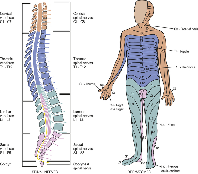
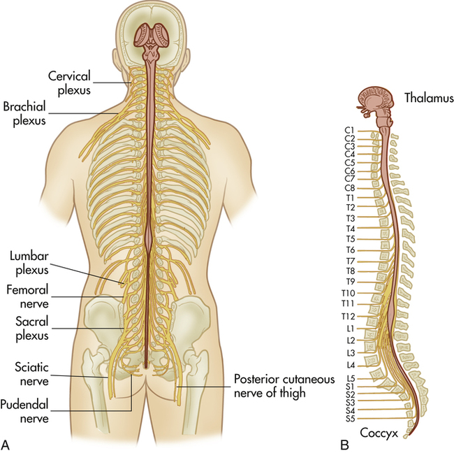
The spinal nerves combine to form the cervical, brachial, lumbar, and sacral plexuses and then innervate the limbs via peripheral nerves (Figure 11-4). Therefore peripheral nerves generally contain fibers from several different spinal nerves. Dermatomes represent the area of skin supplied by a specific spinal nerve and are clinically significant in diagnosing the sensory area of nerve injury (Box 11-2). Nerve injury needs to be distinguished from the cutaneous innervation of the peripheral nerves. A myotome is a muscle or group of muscles supplied by one ventral (motor) nerve. Motor deficits may be attributed to damage in specific myotomes.


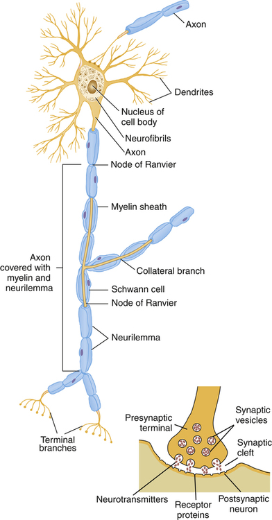
Myelination is a process in which a nerve is enveloped in a myelin sheath. In the peripheral nervous system, this is accomplished by the encircling of a nerve axon by Schwann cells. Gaps between the Schwann cells are called the nodes of Ranvier (Figure 11-5) and expose unmyelinated axons. The significance of this is that nerve conduction in these myelinated nerves is saltatory, jumping from node to node, which increases the conduction velocity.1 This type of myelination ceases just before the interface of the dorsal or ventral nerve root with the spinal cord. At this juncture between the peripheral nervous system (PNS) and the central nervous system (CNS), known as the Obersteiner-Redlich zone, astrocytes and oligodendrocytes form the myelin covering.


Demyelinating conditions such as multiple sclerosis, which affects the CNS, and Guillain-Barré syndrome, which affects both the PNS and CNS, can lead to varying degrees of sensory and motor loss. Remyelination often takes place in the PNS; however, it occurs sluggishly, if at all, in the CNS.


Assessment of the Neurological System


Warning Signs of Neurological Diseases


Assessment of the neurological system begins with the athlete’s clinical history, coupled with listening carefully to the complaints and terms the athlete uses to describe the condition. In general, neurological diseases can present with positive or negative signs or manifestations.


Positive manifestations represent inappropriate excitation of the nervous system. These include hypersensitivity; seizures; movement disorders that include tremor, spasm, and tics; and upper motor neuron signs, such as spasticity, hypertonicity, and hyperreflexia. Some descriptions given by the athlete to indicate positive symptoms include the following: heaviness, weakness, cramps, slow reaction, tiredness, tremors, visual disturbances, incoordination, a deadened feeling, numbness, tingling, or pins and needles. Negative signs or manifestations represent a loss of function. These include paresis; paralysis; hyposensitivity; dementia; aphasia, including receptive-sensory, expressive-motor, and anomic; syncope; neck stiffness; gait dysfunction; movement disorders; incoordination; sensory ataxia or proprioception loss; and lower motor neuron signs including hypotonicity–flaccidity, hyporeflexia, and atrophy. In the case of sport-related concussion (SRC), the history component of the evaluation is crucial in identifying signs and symptoms, and in determining the presence or absence of amnesia and loss of consciousness and evaluating the athlete’s mental status.


The physical examination of the athlete begins with a visual inspection of the spinal column, assessing for deviations, muscular imbalance, or surgical scars. The athlete’s musculature, although typically stronger on the dominant side, should not be particularly unilaterally hypertrophic. The health care provider assesses bilaterally for tremors, atrophy, and muscular tone. The dermatomes are bilaterally assessed for sensation as described in Chapter 2, followed by bilateral comparison of the reflexes. Evaluation of the cranial nerves (described in the next section) and muscular strength tests follow. Muscular strength tests include both range of motion and break tests for the brachial plexus as well as heel and toe walking for the lower extremities. The practitioner notes any weakness or differences and refers the athlete to a physician when a discrepancy occurs in either. Functional knowledge of the myotomes and dermatomes is instrumental in assessment of any neurological issue. The physical examination for SRC should include the assessment of cognition and coordination (balance) along with the aforementioned evaluation of strength, myotomes, and dermatomes.


Clinical neurological signs can assist in differentiating muscle weakness. The pattern of muscle weakness varies depending on the area of injury within the motor unit from nerve cell to muscle: upper motor neuron, lower motor neuron, neuromuscular junction, muscle itself, or manifestation as functional weakness. Upper and lower motor neurons have been previously discussed. Neuromuscular junction disorders are characterized by signs of injury that include fatigable weakness, normal or decreased muscle tone, and normal deep tendon reflexes. Decreased or absent reflexes present with injury at the muscle level (i.e., myopathy). Functional weakness, in contrast to actual weakness, is associated with decreased power in the presence of normal tone, reflexes, and muscle girth.


Understanding the basic anatomy and physiology of the neurological system also helps in identification of injury to higher neurological systems. Cerebral injuries may present with aphasias, apraxias, paresis, paralysis, sensory deficits, and visual and auditory dysfunction. Cerebellar damage leads to varying degrees of ataxia and incoordination, as well as dysmetria and tremors. Neurological injury to the brainstem may manifest with cranial nerve palsies (CN III to CN XII). In addition to these symptoms, many other common signs associated with neurological disease have special terminology (Box 11-3).


BOX 11-3   TERMINOLOGY ASSOCIATED WITH NEUROLOGICAL DISEASES


Agnosia Inability to recognize and interpret sensory stimuli (CVA)


Agraphia Inability to express thoughts in writing (CVA)


Allodynia Sensitivity or pain to nonpainful stimuli (CRPS)


Anisocoria Pupil size inequality of 0.5 to 2.0 mm (neurological disease)


Aphasia Impaired comprehension or expression of written or spoken language (CVA, encephalitis, dysphasia, head trauma)


Apraxia Inability to perform purposeful movements in the absence of weakness, sensory loss, poor coordination, or lack of comprehension (CVA)


Ataxia Incoordination of voluntary movement (CVA, head trauma, GBS, MS)


Aura Sensory or motor phenomenon that indicates the start of a seizure or an impending classical migraine (migraine, seizure)


Babinski’s reflex Abnormal extension of the great toe and extension of the toes with plantar foot stimulation (CVA, head trauma, MS, meningitis)


Brudzinski’s sign Flexion of the hips and knees in response to passive flexion of the neck (meningitis, trauma, SAH)


Decerebrate rigidity Abnormal extensor responses in the upper and lower limbs (cerebral trauma)


Decorticate rigidity Abnormal flexor response in the upper extremity and extensor response in the lower extremity (CVA, head trauma)


Deep tendon reflexes Abnormally brisk muscle contraction with tapping; hyperactive on the muscle tendon (CVA, MS); abnormally slow muscle contraction with tapping; hypoactive on the muscle tendon (GBS)


Diplopia Double vision (CVA, encephalitis, MS, head trauma, migraine)


Drooling Loss of saliva from the mouth (CVA, GBS, seizure)


Dysarthria Impaired articulation (CVA, MS)


Dysdiadochokinesia Difficulty performing rapidly alternating movements (CVA—cerebellar)


Fasciculations Irregular contractions of groups of muscle fibers (GBS)


Footdrop Plantar flexion of the foot caused by impaired ability to dorsiflex the foot (CVA, MS, GBS)


Gait, spastic Abnormal gait with extended stiff legs, causing dragging of the foot (MS, CVA)


Gait, steppage with footdrop Abnormal gait with exaggerated hip and knee flexion to clear the dragging toes (GBS, MS)


Headache Pain in the head, retroorbital, and cervical areas


Hemianopia Loss of vision in one half of the visual field (CVA)


Hyperesthesia Increased sensitivity to touch, pain, and temperature (CVA, CRPS)


Incontinence Involuntary loss of urine or feces (CVA, MS, head trauma)


Kernig’s sign With a patient in the supine position and leg flexion, there is resistance and pain with leg extension (meningitis, SAH)


Level of consciousness Reduced alertness ranging from lethargy to stupor (CVA, encephalitis, head trauma—bleeds, seizure, meningitis)


Light flashes Bright stars, streaks, or spots in the visual field (migraine, head trauma)


Miosis Unilateral constriction of a pupil (cluster headache)


Muscle atrophy Wasting of muscle size (CVA, MS)


Muscle flaccidity Profound weakness with lack of active muscle movement and resistance (CVA, GBS, seizures)


Muscle spasticity Increased resistance to movement (CVA, MS, head trauma)


Muscle weakness Reduced muscle strength (CVA, MS, head trauma, GBS, seizure)


Myoclonus Spasms of a muscle or group of muscles (viral encephalitis)


Nuchal rigidity Profound stiffness of the neck with flexion (meningitis, encephalitis, head trauma—bleeds)


Nystagmus Involuntary oscillations of one or both eyes (CVA, MS, encephalitis, head trauma)


Ocular deviation Abnormal movement of one or both eyes (CVA, MS, encephalitis, head trauma, meningitis)


Paralysis Complete loss of voluntary movement (CVA, encephalitis, MS, migraine, seizure, head trauma—bleeds)


Paresthesia Abnormal sensations in the distribution of peripheral nerves (CVA, GBS, migraine, MS, head trauma, seizure)


Photophobia Increased sensitivity to light (meningitis, migraine)


Ptosis Drooping of the upper eyelid (migraine)


Pupils, nonreactive Absence of constrictive response of the pupil to light (encephalitis)


Pupils, sluggish Abnormally slow constrictive response of the pupils to light (encephalitis, MS)


Rhinorrhea Loss of thin mucus from the nasal passages (cluster headache)


Romberg’s sign Impaired balance with eyes closed and feet together (MS)


Scotoma Focal area of darkness or blindness in the visual field (migraine)


Seizure Abnormal cerebral electrical activity presenting disturbances (CVA, MS, head trauma, with motor, sensory, autonomic, or psychic encephalitis)


Tremors Rhythmical shaking of an extremity from involuntary contraction and relaxation of opposing muscles (MS)


Vision, blurred Impaired visual acuity (CVA, MS, migraine, concussion)


Vision, lost Inability to sense visual stimuli (concussion)


CRPS, Complex regional pain syndrome; CVA, cerebrovascular accident; GBS, Guillain-Barré syndrome; MS, multiple sclerosis; SAH, subarachnoid hemorrhage.


Neurological presentations can range from emergent, to urgent, to routine in severity and in their need for medical evaluation and management. The box entitled “Red Flags for Urgent Intervention” provides a framework of warning signs and symptoms that warrant immediate referral to a physician or an emergency department. Indications for referral of specific disorders are addressed in the section Pathological Conditions.


Cranial Nerves and Cranial Nerve Testing


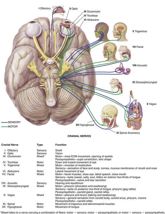
The cranial nerves emerge from the cranium, as opposed to spinal nerves, which emerge from the spinal cord (Figure 11-6). The cranial nerves provide sensory and motor innervation to the head and neck, including voluntary as well as involuntary muscle function, and sensation (see Table 2-4, Cranial Nerve Function). Testing these nerves is essential to ascertaining their integrity as well as noting discrepancies that may indicate a medical condition.


The olfactory nerve (CN I) can be tested by placing different-smelling substances underneath a single nostril with the other nostril occluded. Both nares are tested because injuries to this


nerve are usually unilateral. The clinical spectrum of pathology can range from normal function to anosmia, in which the patient can discern only ammonia, to organic dysfunction, in which the patient cannot recognize any smells.


The optic nerve (CN II) carries visual information within the complex visual system. The fibers of the optic nerve carrying information from the right half of the retina cross over and join with those same fibers of the contralateral optic nerve. Together, these fibers form the optic tract. The optic tract then forms optic radiations that eventually synapse on the primary visual cortex. Different clinical visual defects will occur depending on the area of lesion within the optic pathway.1 Complete assessment of the optic nerve requires testing the visual fields, acuity, and pupillary light reflex.


To test the visual fields, the health care provider faces the athlete, who is looking straight ahead. The athletic trainer moves the fingers of one hand within the athlete’s peripheral vision and monitors the athlete’s response about which finger is moving. Next, the practitioner focuses on individual eye deficits by repeating this test while the athlete closes one eye. The Snellen eye chart, which is described in Chapter 12 is used to test visual acuity. Alternatively, the athlete may be asked to read something.


The oculomotor nerve (CN III) controls the pupillary light reflex. When a light is shone in an athlete’s eye, the pupil normally constricts (direct response); the pupil of the other, unstimulated eye should constrict as well (consensual response). Any deviation from this is abnormal. This nerve is also responsible for eye adduction (toward the midline) and downward movement.


The trochlear (CN IV) and abducens (CN VI) nerves can also be tested by monitoring the movements of the extraocular muscles. The athlete is asked to visually follow a pen as it is slowly moved within the visual field. Then the athletic trainer passes the pen across the midline space toward the athlete’s nose, watching for movement of both eyes in toward the midline and testing for convergence. Saccadic eye movements are tested by having the athlete look in each direction and watching the coordination and quality of movement. Specifically, the trochlear nerve (CN IV) is responsible for upward eye movement and the abducens nerve (CN VI) coordinates eye movement laterally away from the nose (Figure 11-7).


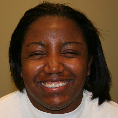
The health care provider tests response of the trigeminal nerve (CN V) and the facial nerve (CN VII) by observing for symmetry when the athlete bares teeth, whistles, looks up at the ceiling, and clenches eyes closed (Figure 11-8). Sensory testing of the trigeminal nerve is performed with light touch and pinprick in the divisions of the trigeminal nerve on both sides. If any abnormalities are found, further tests are warranted. When testing the motor function of the trigeminal nerve, the athletic trainer has the athlete clench teeth while placing a hand under the athlete’s chin to resist jaw opening. Any noticeable muscle atrophy or elicited muscle weakness indicates an abnormal test. Sensory testing of CN VII as well as CN IX (glossopharyngeal) appraises the athlete’s ability to distinguish taste. For example, the athlete might be asked to tell the difference between sweet and sour.


Testing of the vestibulocochlear nerve (CN VIII) involves auditory testing of hearing and equilibrium. Auditory testing is performed using the Rinne test and the Weber test (see Box 13-2). In both tests a vibrating tuning fork is placed at various points on the patient’s head and the patient is asked to identify which placement is louder. Conductive deafness occurs when conduction of sound is impaired, as opposed to sensorineural deafness, in which there is neurological disruption. Auditory testing may also be conducted by creating a sound—such as snapping fingers—behind each of the athlete’s ears and looking for a response.


Tests of the glossopharyngeal (CN IX), vagus (CN X), and hypoglossal (CN XII) motor nerves involve observing the athlete’s tongue and mouth anatomy and watching for deviations in tongue movement when the mouth is opened and the tongue stuck out. The athlete is asked to push tongue against cheek or a depressor to demonstrate strength. The athletic trainer watches the uvula for deviation from midline as the patient says “aah.” The vagus nerve (CN X) controls the gag reflex, which is assessed by touching each side of the pharyngeal wall behind the tonsils and observing movement of the uvula. The vagus nerve is also responsible for movement of the larynx and pharynx, which can be tested together by observing the quality and coordination of movement while the athlete swallows water.


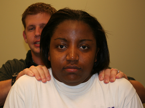
The athletic trainer tests the spinal accessory nerve (CN XI) by focused examination of the sternocleidomastoid and trapezius muscles on both sides of the body. The strength of the trapezius muscle is assessed through a resisted shoulder shrug (Figure 11-9), and the sternocleidomastoid muscle is assessed by resisting both turning and lifting of the chin. The presence of atrophy or weakness during resisted muscle movement suggests nerve injury.


Pathological Conditions


Pathological conditions discussed in this chapter are divided into sections as follows; brain and spinal cord disorders (sport-related concussion, stroke, Guillain-Barré syndrome); paroxysmal disorders (headache, seizures); neuromuscular disorders (multiple sclerosis [MS], amyotrophic lateral sclerosis [ALS]); and pain disorders (complex regional pain syndrome).


The presentation and diagnosis of many of neurological conditions are based on a process of exclusion. The physician must exclude all other possible explanations for the symptoms in order to arrive at the correct diagnosis. Conditions such as Guillain-Barré syndrome, MS, ALS, and complex regional pain syndrome all have a rather insidious onset, and no single diagnostic test confirms the presence of the affliction. A thorough history is paramount in helping the clinician arrive at the correct diagnosis. Patients often dismiss symptoms and attribute them to fatigue (vision problems, balance issues, headaches), and a complete medical history should illuminate the symptoms specific to the neurological condition.


Brain and Spinal Cord Disorders


Sport-Related Concussion


Sport-related concussion (SRC) is a common injury in recreational activities and sports that may have possible short- and long-term sequelae. Reports have estimated that approximately 1.6 to 3.8 million SRCs occur annually.1 It is also speculated that these numbers may be higher due to those SRCs that are not recognized by the athlete, and thus not reported to medical personnel.24 Data from two national injury surveillance systems found that concussion represented 8.9% of all high school and 5.8% of all collegiate athletic injuries.5


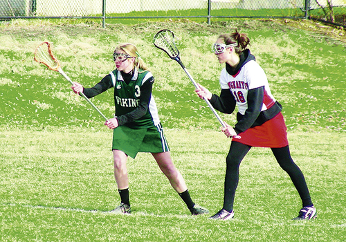
An evidence-based definition was published after the Third International Conference on Concussion in Sport, held in 2008.6 This definition, provided in Box 11-4, is more comprehensive in nature and identifies clinical, pathological, and biomechanical features of concussion that are important in identifying these injuries.6 It was found that an unacceptably high percentage of high school players were playing with residual symptoms from a prior head injury7 (Figure 11-10).


Signs and Symptoms

The health care provider should be able to identify signs and symptoms associated with SRC. These signs and symptoms are often categorized as somatic (physical), emotional, cognitive, and sleep-related.8 Box 11-5 lists the specific symptoms that warrant referral to a medical facility. Each athlete may present differently with respect to the severity and duration of reported symptoms. In a National Collegiate Athletic Association (NCAA) concussion study,9 there was an increase in reported symptoms immediately after concussion, followed by a gradual decrease in symptom complaints throughout the first week, with 91% of athletes returning to their baseline symptom levels by day 7 postinjury. Headache is the most common symptom reported and tends to last the longest.1012


BOX 11-5   PHYSICIAN REFERRAL CHECKLIST


Day-of-Injury Referral


1. Loss of consciousness on the field


2. Amnesia lasting longer than 15 minutes


3. Deterioration of neurological function


4. Decreasing levels of consciousness;


5. Decrease or irregularity in respirations


6. Decrease or irregularity in pulse


7. Increase in blood pressure


8. Unequal, dilated, or unreactive pupils


9. Cranial nerve deficits


10. Any signs or symptoms of associated injuries (spine or skull fracture, bleeding)


11. Mental status changes (lethargy, confusion, difficulty maintaining arousal, confusion, or agitation)


12. Seizure activity


13. Vomiting


14. Motor deficits subsequent to the initial on-field assessment


15. Sensory deficits subsequent to the initial on-field assessment


16. Balance deficits subsequent to the initial on-field assessment


17. Cranial nerve deficits subsequent to the initial on-field assessment


18. Postconcussion symptoms that worsen


19. Additional postconcussion symptoms as compared with what was reported on the field


20. Symptoms that do not improve by the end of the game/practice (especially at the high school level or younger)


Requires immediate transportation to nearest medical facility


From Guskiewicz KM, Bruce SL, Cantu RC, et al: National Athletic Trainers’ Association Pronouncement Committee: position statement on sport-related concussion, J Ath Train 39:280-297, 2004.


Differential Diagnosis and Referral

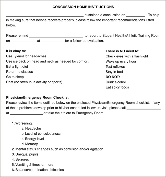
As part of the initial assessment of SRC, the health care provider should attempt to rule out more severe head injuries including skull fracture, cerebral contusion, and epidural hematomas.6,7,13,14 Assessment of these injuries may reveal acute localized swelling, deformity, prolonged loss of consciousness (LOC), irretractable vomiting, and frequently, multiple positive neurological examination findings such as cranial nerve abnormalities and motor or sensory weakness. Positive findings during an initial examination for one or more of these indications should warrant immediate transfer to an emergency department capable of managing a neurosurgical emergency.13 In addition, the clinician should continue to monitor the athlete for any signs of altering or deteriorating condition in the acute injury phase and throughout the first week postinjury that would increase suspicion of a subdural hematoma. Patients should be given a home instruction form that lists the red flag warnings for immediate referral and other information about home care of the SRC (Figure 11-11).


Physician referral is also indicated for athletes who are not experiencing a typical recovery, for those who may still be symptomatic in the weeks after their initial injury, and for those who demonstrate an increase in symptoms in the postacute phase. At this time, referral to a specific specialty may be necessary. For example, an athlete with sleep disturbances may benefit from a referral to a neurologist, whereas an individual with cognitive difficulties may be best suited for a referral to a neuropsychologist.13


Diagnostic Tests

Because SRC is a functional injury and does not normally include structural injury to the brain, most diagnostic tests will result in negative imaging findings, thus making neuroimaging of little value for most SRCs.6,7,13 Suspicion of an intracranial hemorrhage or hematoma should result in immediate referral for a computed tomography (CT) scan, Functional magnetic resonance imaging (MRI), or other imaging study. Moreover, the sudden presence or deterioration of symptoms that had previously resolved or had remained stable necessitates neuroimaging for a subdural hematoma, which may not be noted on a CT scan or MRI for 1 to 2 weeks after the initial concussion.13 However, it is imperative to understand that a negative imaging finding does not rule out an SRC and should not be used to prematurely return a patient to activity.


Assessment and Management

The assessment of SRC should begin off-season or during preseason in conjunction with the preparticipation physical examination (PPE) to obtain an adequate concussion history and to assess baseline measures of symptom reports, postural stability, and neurocognition. The PPE should include a thorough neurological injury history, including a history of sport-related concussion and other concussive injuries (e.g., motor vehicle accidents, falls).15,16 The PPE should contain an adequate series of questions regarding concussion history, including queries about perceived previous concussions and those focusing on earlier concussion-related symptoms sustained during both sport and nonsport activity.6,15,17 Knowledge of an athlete’s concussion history is important as research has demonstrated a relationship between previous concussions and increased risk for sustaining subsequent injuries.1820 In addition, baseline measures of symptoms, neurocognitive function, and postural stability should be obtained from the athlete when he/she is in a healthy state for future comparison with postconcussion scores.


After a concussion, the athlete should be assessed immediately for symptom reports, mental status, and postural stability. Serial assessments using the tools described below should take place at planned intervals (postgame, 24 and 48 h postinjury) to assess the athlete’s recovery (Box 11-6). Once the athlete reports that he/she is symptom free, the more complex neurocognitive assessments should be administered to determine whether cognitive recovery has occurred as well. Scores on all assessment tools should meet or improve on baseline scores before a return-to-play progression is begun.


BOX 11-6   GRADED SYMPTOM SCALE


A graded symptom scale (GSS), like the one shown here, is a tool used to determine progress after a concussion. Serial assessments, such as for postconcussive signs and symptoms (PCSS), are often indicated by marking “yes” or “no” in the various categories.


Sep 3, 2016 | Posted by in MUSCULOSKELETAL MEDICINE | Comments Off on Neurological System

Full access? Get Clinical Tree

Get Clinical Tree app for offline access