Figure 15.1
Open ended hair-pin arrangement of metaphyseal blood vessels adjacent to the physis with transphyseal vessels crossing across into the epiphysis. This arrangement predisposes infants to septic arthritis and growth plate injury. The transphyseal vessels disappear after the first year of life and the physis becomes a barrier to the haematogenous spread of infection from the metaphysis to the epiphysis. (Modified from Benson et al Children’s Orthopaedics and Fractures, Springer, 2009)
2.
Chronic & Subacute Osteomyelitis
Organisms of low virulence may proliferate in patients with an impaired host resistance to cause a subacute or chronic osteomyelitis. In subacute osteomyelitis, a walled off, pus-filled, cavity (i.e. bone abscess or Brodie’s abscess) may form. This is usually found in sites of rapid skeletal growth, such as the proximal tibia, distal femur, humeral metaphysis or distal radius. Symptomatically, patients tend to have only mild and intermittent pain in the absence of a systemic inflammatory response.
Chronic osteomyelitis results from an untreated or inadequately treated acute infection, or it may silently bypass the “acute” phase and first come to light after the development of sequestrum, involucrum and a sinus tract.
Pathophysiology of Septic Arthritis
1.
Bacterial Seeding
This can occur in a number of ways:
1.
Haematogenous spread to synovial capillaries, which form micro-abscesses that rupture into the joint.
2.
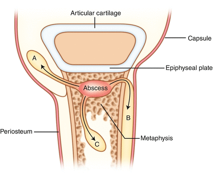
Transphyseal vessels present in young infants can permit the spread of bacteria from a metaphyseal osteomyelitic focus into the epiphysis, which can perforate the articular cartilage to enter the joint. The osteomyelitic lesion often becomes apparent several days after the initial presentation of an acute episode of septic arthritis (Fig. 15.2).


Figure 15.2
Showing the spread of pus from a metaphyseal focus of osteomyelitis – (A) directly into the joint, (B) into a subperiosteal abscess, (C) into the medullary canal. (Modified from Benson et al Children’s Orthopaedics and Fractures, Springer, 2009)
3.
Direct inoculation into the joint via a periarticular abscess or trauma.
2.
Local Inflammation
An acute synovitic reaction leads to the development of a seropurulent exudate, resulting in a painful joint effusion, which can cause joint subluxation or even dislocation in severe cases. Pus may even burst out of a joint to cause an abscess.
3.
Articular Erosion
The presence of bacterial and proteolytic enzymes released by leucocytes leads to a progressive and irreversible erosion of the articular cartilage, and the largely cartilaginous epiphysis.
4.
Epiphyseal Avascular Necrosis
Increasing intra-articular pressure further reduces perfusion of the remaining epiphysis, causing avascular necrosis if left untreated.
5.
Healing phase
Depending on the timing and success of medical intervention, complete resolution of the infection with a return to normal function can occur. However, delay in treatment may lead to partial loss of articular cartilage with fibrosis of the joint, bony ankylosis and eventual bone destruction, leaving the child with a lasting deformity.
Pathogens Causing Infection
A host of organisms can cause either osteomyelitis or a septic arthritis. Normally between 50% and 75% of organisms are identified. They include:
Staphylococci – These gram + ve cocci are responsible for the majority of infections. Staphylococcus aureus is a constituent of normal skin flora. Many strains are possible and resistance to antibiotics common.
MRSA – Methicillin Resistant Staphylococcus Aureus is becoming more common and isolated more frequently. It is more common amongst those with risk factors and institutionalised patients.
Streptococci – Another of the Gram + ve family, the beta haemolytic strain accounts for most cases. It tends to effect neonates or older children.
Haemophilus influenzae – Particularly in populations which do not have an immunization program against it.
Others – Escherichia coli, Kingella kingae and mycobacteria.
Symptoms and Signs
As haematogenous spread is usually the commonest cause of septic arthritis in this group of patients, special attention should be made to investigate for and treat any distant sites of primary infection. The source of infection could arise from a multitude of sites, including the CNS, chest, abdomen, spine, upper respiratory tract, ears, urinary tract and skin, including the umbilical cord or intravenous cannula sites, hence the extra vigilance required when assessing such patients (Table 15.1).
The clinical features of paediatric musculoskeletal infection differ according to age and site.
Table 15.1
Risk factors for the development of musculoskeletal infection
Malnutrition |
Sickle cell disease |
Immunosuppressive drugs |
Diabetes |
Immunocompromised states (e.g. HIV infection, Chemotherapy, Steroid use) |
Neonates |
Postoperative |
1.
Neonates (up to 4 weeks old):
Newborns tend to have an atypical presentation with few clinical signs. Constitutional symptoms include general irritability and a refusal to feed. This can be associated with or without signs of a systemic inflammatory response syndrome (SIRS). Careful inspection, palpation and movement of the affected limb and/or joint should take account any local inflammatory features, such as subtle signs of effusion (abnormal skin creases; bulge over joint), pain, warmth and resistance to movement.
2.


Infants (1–12 months) and children
The more typical features of septic arthritis include inability of the patient to bear weight on the affected joint or actively move it (pseudoparalysis) due to the presence of a tense joint effusion and pain. Passive movement of the joint helps to differentiate septic arthritis from the pain caused by osteomyelitis, which does not usually result in painful passive joint movements. There may be overt signs of inflammation, including warmth to touch, swelling and a hyperaemic appearance of the overlying skin. As with neonates, associated signs of a SIRS or sepsis may be present, requiring careful monitoring of the pulse, temperature and hydration status.
Stay updated, free articles. Join our Telegram channel

Full access? Get Clinical Tree


