Figure 8.1
Diagram demonstrating the displacement of the proximal femoral epiphysis and subsequent rotation of the femoral neck relative to the head. (a) Normal, (b) mild, (c) moderate, and (d) severe
Epidemiology
SCFE is the most frequent hip disorder amongst adolescents and occurs during the adolescent growth spurt. Males are more commonly affected than females by a ratio of 3.2:1 Incidence rates are between 2 and 10 in 100,000. At time of presentation, most boys are aged between 12 and 15 years and most girls between 10 and 13 years. Obesity is a well-defined feature of SCFE with over half of affected children weighing over the 95 centile for their age group. There are significant differences in incidence among differing races, with African Americans and Polynesians having the highest rates.
In unilateral cases, the left hip is more commonly affected. Up to 20% of cases will be bilateral at presentation. Of the 80% unilateral cases, 20–40% will develop a symptomatic slip of the contralateral side during adolescence, the majority if these occurring within 18 months from diagnosis of the initial slip.
Aetiology
The exact aetiology of SCFE is unclear. Local anatomical and perhaps hormonal changes cause mechanical instability of the proximal femoral physis with inability to resist load across it. The failure of the physis across the hypertrophic zone results in the slippage.
There are numerous conditions associated with SCFE and the susceptible physis. These include:
Endocrine factors:
The most common endocrine conditions in children with SCFE are:
Hypothyroidism.
Panhypopituitarism.
Growth hormone abnormalities.
Hypogonadism.
Other endocrine causes include:
Hyperparathyroidism.
Renal osteodystrophy.
Up to eightfold increase in risk for SUFE.
Due to secondary hyperparathyroidism.
Mechanical factors.
Femoral neck retroversion.
Decreased femoral neck-shaft angle.
Superior acetabular retroversion and increased superolateral femoral head coverage.
Increased acetabular depth.
Syndromic factors.
Trisomy 21 (Down’s syndrome).
Pathogenesis
Histological analyses of the physis in SCFE suggest general disorganisation of the growth plate, with increased chrondroblast cell turnover (apoptosis). There is clear abnormality within the hypertrophic zone, which is found to be wider than the resting zone, with a reduced amount of collagen with poor orientation and poor columnar organisation of the cells.
Symptoms and Signs
Onset of symptoms may be sudden or, more commonly, gradually develop over the preceding weeks or months. Typically, the patient will complain of pain in the hip or groin. Thigh pain is also a common symptom and up to 25% of children may present with knee pain.
On examination, the child may walk with an antalgic or Trendelenburg gait, with the affected limb in external rotation. There is increasing limitation of hip movements with increasing severity of SCFE. Obligatory external rotation on passive hip flexion (Drennan’s sign), with an associated loss of internal rotation is a common finding. Limb shortening is seen in severe cases. Patients with an unstable slip demonstrate complete inability to bear weight even with support and often are reluctant to move the limb.

Investigations
Plain radiographs of both hips are the standard imaging modality in SCFE. Anteroposterior (AP) and lateral radiographs are sufficient to confirm the diagnosis. The earliest findings can be seen on lateral radiographs.
Radiographic features on AP (Fig. 8.2) or shoot through/frog lateral (Fig. 8.4) radiographs include:
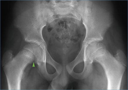
Figure 8.2
AP radiograph showing Klein’s line and the metaphyseal blanch sign, arrowhead
1.
Widening and irregularity of the physis.
2.
Decreased height of the capital femoral epiphysis (seen when the epiphysis lies behind the femoral neck).
3.
Metaphyseal blanch sign – a dense crescent shaped area caused by the superimposed displaced epiphysis (arrow Fig. 8.2).
4.
Callus formation in chronic slips.
5.
Part of the femoral epiphysis normally lies lateral to Klein’s line, a line drawn along the lateral aspect of the femoral neck. In SCFE, the line does not pass through the femoral head; this is known as Trethowan’s sign. Comparison with the contralateral side may be useful (Figs. 8.2 and 8.3).
Figure 8.3
(a, b) Klein’s line in (a) normal and (b) SCFE (Reproduced from Houghton, KM, Review for the generalist: evaluation of pediatric hip pain, Pediatric Rheumatology, 2009 7:10)
Features on lateral radiographs include:
1.
Posterior step off and slipping of the epiphysis, or posterior callus (Fig. 8.4).
Figure 8.4
Frog leg lateral radiograph of a bilateral SCFE
2.
Southwick angle (Fig. 8.5).
Figure 8.5
Diagrammatic representation of the Southwick angle. a Line connecting anterior and posterior margins of the physis, b line perpendicular to a, c line marking the centre of femoral neck and shaft
The epiphyseal-shaft angle of the affected side subtracted from the normal side.
The normal epiphyseal shaft angle is around 10–12°.
Computed tomography (CT) has limited benefit in SCFE. Situations where it is useful, includes:
In late presentations of SUFE, to assess the degree of physeal closure. If closed, correction can generally only be safely achieved with extracapsular osteotomies in order to avoid avascular necrosis.
In the presence of femoral head collapse, secondary to avascular necrosis, to assess penetration of metalwork in to the joint.Related posts:

Stay updated, free articles. Join our Telegram channel

Full access? Get Clinical Tree


