Figure 10.1
Langenskiold’s radiographic classification of infantile Blount disease. Progressive stages from mild medial epiphyseal-metaphyseal beaking (stage I) to complete medial proximal tibial physeal arrest (stage VI) (Reproduced with permission from Tachdjian’s pediatric orthopaedics 4th Edition, 2nd volume)
The metaphyseal-diaphyseal angle (MDA) or Drennan’s angle assessed on an AP radiograph can be used to differentiate between physiological varus and infantile Blount’s disease (Figs. 10.2 and 10.3).
Figure 10.2
The tibial metaphyseal-diaphyseal angle (MDA), created by the intersection of a line connecting the most prominent medial portion of the proximal tibial metaphysis (the “beak”) and the most prominent lateral point of the metaphysis with a line drawn perpendicular to the long axis of the tibial diaphysis
Figure 10.3
Long leg radiograph showing bilateral Blount’s disease
Angles <11° suggests physiological genu varum.
Angles of >16° suggests Blount’s disease.
Those in between with medial beaking are more likely to progress to true infantile Blount’s disease and should be closely monitored.
Management
A simple algorithm for treatment is as follows:
Patients ≤3 years. who are Langenskiold’s stage I/II can be initially treated conservatively in a long leg anti-varus brace during the day.
Those ≥4 years, with progressive radiographic deformity or Langenskiold’s stage III–IV are likely to require surgery.
Surgical options include the following:
High tibial osteotomy – The goal is correct the varus, flexion, and internal rotational deformities of the tibia. By reducing the weight bearing across the medial joint, growth and subsequent correction of the deformity ensues. The younger the patient, the greater the risk of recurrence, hence overcorrection is common. Correction can be achieved with acute or staged osteotomies and with immediate internal fixation or treatment with a frame. An oblique osteotomy can be used to correct both the varus and internal rotation components of the deformity (Fig. 10.4).
Figure 10.4
Radiographs show reoccurrence after high tibial osteotomy
In severe cases, a medial hemiplateau elevation osteotomy may be required.
The use of guided growth plates can be used to perform a hemiephysiodesis/growth modulation. Satisfactory results have been observed with this technique, although metalwork failure remains an issue.
Complications
Possible complications from surgery include the following:
Compartment syndrome.
Nerve injury (common peroneal nerve).
Infection.
Malunion/nonunion.
Recurrence.
Physeal arrest.
Late Blount’s Disease
The less common and less severe adolescent form of Blount’s affects children over the age of 10 and is typically associated with obesity. Excess weight is thought to cause overloading of the medial joint on an already mildly varus knee (Fig. 10.5).
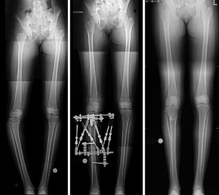
Figure 10.5
Long leg radiographs showing an late Blount’s deformity (left), corrected using a frame (middle) and post-treatment alignment (right)
Features associated with the adolescent form include:
A greater chance of unilateral cases, possibly increasing limb length discrepancy.
Distal femoral physeal growth disturbances, exacerbating the varus deformity.
Typical radiographic features include:
Associated femoral deformity (usually valgus).
Widening of the medial tibial physis.
Widening of the lateral femoral physis.
Narrowing of the tibial epiphysis.
Beaking is not as common as the infantile type.
As with infantile Blount’s, the MDA is greater than 16°.
Management
In untreated cases, deformity worsens and early osteoarthritis sets in due to altered joint kinematics. Reversal of the driving force (obesity) should be addressed but as there is less potential for remodeling due to age, non-operative treatment is of limited value and poor outcomes are typically seen in all but mild cases.
As with infantile Blount’s, surgery typically involves correction via a proximal tibial osteotomy. Once again, this can be acute or gradual and fixed acutely or using a frame. Overcorrection is not required at this advanced age, as ensuing growth tends to be along the mechanical axis. In some patients with enough remaining growth potential, hemiepiphysiodesis remains an option. Treatment of femoral deformity by similar methods may also be considered.
Recurrence of deformity following a high tibial osteotomy implies severe and possibly irreversible medial proximal tibial physeal growth disturbances. In these cases, future treatment has to be individualized taking into account the age, deformity, joint distortion and leg length inequality.
Focal Fibrocartilaginous Dysplasia (FFCD)
FFCD is a rare and benign condition affecting the proximal tibia of toddlers and infants (Fig. 10.6). It has the same prevalence in both sexes. The clinical features of tibial FFCD include:
Painless unilateral tibia vara.
Medial tibial torsion.
Leg length discrepancy.
Limping or normal walking.
Figure 10.6
A radiograph showing left sided tibia vara as a result of Focal Fibrocartilaginous Dysplasia
It is a result of failure of differentiation of the mesenchymal anlage in the area of the pes anserinus. Persistence of a fibrocartilage focus hampers growth on the medial aspect of proximal tibia. Given the physis is normal, the deformity typically corrects. In the rare cases where it doesn’t, correction may be achieved by curettage of the lesion.
Congenital Dislocation of the Knee (CDK)
Congenital dislocation of the knee or “back knee” comprises of a spectrum of deformities ranging from subluxation to complete dislocation. The incidence is estimated at 1 per 100,00 live births.
Related posts:

Stay updated, free articles. Join our Telegram channel

Full access? Get Clinical Tree


