Symptoms/Signs
The signs present will depend on the age of the child being examined. The key for examination is to identify if the hip is dislocated, or if not, whether it is unstable.
In unilateral dislocation, asymmetrical reduction of hip abduction can be useful to identify abnormality, although this may be less helpful in the bilateral dislocation. Thigh skin crease asymmetry can be identified, although this is neither sensitive nor specific, with 1 in 4 children demonstrating some asymmetry (Fig. 6.1). Widening of the perineum may be noted on the affected side.
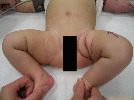
Figure 6.1
Photograph demonstrating asymmetry of thigh creases in left sided DDH
Leg lengths can be examined with the heels and knees together, looking for relative shortening of the femur (Galeazzi’s test). This will demonstrate any cause for femoral shortening (e.g. proximal femoral focal deficiency) (see Chap. 13) and is not specific.
The widely used tests for screening in the neonate are the Barlow’s and Ortolani’s test. These tests are performed as part of a neonatal check and again at 6 weeks in the UK. The neonate is examined with the nappy removed, supine on a firm examination couch. The hips and knees are flexed to 90° and the hands placed over each thigh, with the middle finger resting on the greater trochanter and the thumb over the medial thigh.
Ortolani’s test (Fig. 6.2) involves gentle abduction of both thighs combined with an elevation force applied by the middle finger on the greater trochanter. In the normal infant, full abduction to 90° should be achieved. If the hip is dislocated, then there will be reduced hip abduction and there may be a “clunk” of relocation as the femoral head relocates, moving over the posterior acetabular wall. If the hip is initially enlocated, or is dislocated and irreducible, this will not occur.
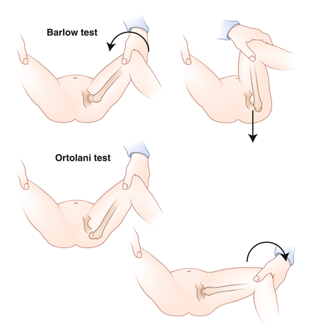
Figure 6.2
Diagram demonstrating the Barlow and Ortolani tests
Barlow’s test (Fig. 6.2) demonstrates instability of a reduced hip. With the hands in the same position as previously, the hips are held flexed to 90° and adducted. A posteriorly directed force is applied while feeling for the femoral head dislocating posteriorly with the middle fingers. Once again a “clunk” may be appreciated suggesting a dislocatable hip.
Both of these tests become less useful as the child grows older; by 3 months the increased muscle tone and stiffness prevent easy dislocation/relocation. For later diagnosis, the most sensitive test is measurement of abduction in 90° of flexion. The affected hip will demonstrate reduced abduction in comparison to the normal side.
In the walking child, a Trendelenburg gait may be observed, reflecting the defunctioning of the abductors. The child may walk on their tip-toes in view of the shortening on the affected side. The bilateral dislocation presenting at walking age demonstrates a “waddling” gait, often with an associated hyperlordosis of the lumbar spine.
Investigations
Imaging modality used is dependent on the age of the infant.
Ultrasound
Ultrasound is useful in the neonate with little ossification of the acetabulum and no ossification centre of the femoral head (<3 months). The use of ultrasound was initially introduced by Graf in the 1980s, using the method to classify the hip according to bony and cartilaginous morphology as viewed on a coronal section (Fig. 6.3a–d). The alpha angle is calculated according to the angle of coverage of the bony acetabular roof, the beta angle representing the cartilaginous roof and labrum. This method requires a reduced hip to produce accurate measurement (Table 6.1).
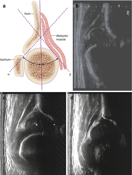
Figure 6.3
(a) Coronal section of hip demonstrating alpha and beta angles (reproduced with permission from Tachdjian’s Pediatric Orthopaedics, ed 4. Philadelphia. WB Saunders). (b) Sonogram illustrating Graf IV (dislocated hip). (c) Sonogram illustrating a Graf IIc (alpha angle 48°) Hip at 6 weeks of age. Decision to treat with Pavlik Harness. (d) Sonogram of Hip (3c) after 6 weeks of Pavlik harness treatment, demonstrating normal acetabular morphology
Table 6.1
Sonographic classification according to Graf
Graf type | Alpha angle | Beta angle | Comment | Treatment |
|---|---|---|---|---|
Ia | >60 | <55 | Normal hip | None |
II* | 50–59 | Physiologically immature (<12 weeks) | Observation | |
IIa | 50–59 | <77 | Shallow acetabulum (>12 weeks) | Pavlik Harness |
IIb | 50–59 | <77 | Delay of ossification of femoral head | Pavlik Harness |
IIc | 43–49 | <77 | Acetabular deficiency | Pavlik Harness |
IId | 43–49 | >77 | Everted cartilage roof/labrum | Pavlik Harness |
III | <43 | Subluxed hip | Pavlik Harness | |
IV | Not measurable | Dislocated hip | Trial of reduction with Pavlik Harness |
Dynamic methods have been demonstrated by Harcke, in which the Barlow and Ortolani tests are performed under ultrasound guidance. This provides information of stability, rather than morphology. The method is dependent on the skill and experience of the sonographer. Rosendahl described use of a combination of dynamic and static methods to allow the identification of both morphology and stability.
Plain Radiographs
Anteroposterior views of the pelvis and hips may be more useful from 3 months of age. Various lines have been described to aid in the assessment of the paediatric radiograph (Fig. 6.4):
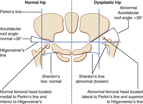
Figure 6.4
Demonstration of the radiographic parameters seen in the normal and dislocated hip
Shenton’s line- A line following the medial neck of the femur should continue in an unbroken curve to follow the inferior edge of the pubis. This should be intact in any view of the pelvis; if disrupted then dislocation or subluxation should be suspected.
Hilgenreiner’s and Perkin’s lines – A straight horizontal line drawn between the triradiate cartilages in both hips. This can be used in conjunction with Perkin’s line, which is a vertical line drawn down from the lateral edge of the acetabular sourcil, perpendicular to that of Hilgenreiner’s line. The normal femoral head should lie in the inferior and medial quadrant produced by these two lines.
Acetabular index- the angle between Hilgenreiner’s line and the slope of the acetabular sourcil forms the acetabular index. This should measure <30° at birth, falling to <20° by 2 years.
Other measurable parameters include Sharp’s Angle and the Centre edge angle of Wiberg (Fig. 6.5) Measurement of radiographic parameters in the child should be taken with caution. Inter and intraobserver variability has been noted for many of these measurements, not to mention the difficulty of accurately positioning the patient to prevent rotated images. The trend of measurements over time is more valuable than the measurements made on a singe radiograph.
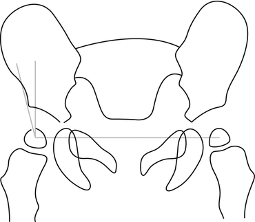
Figure 6.5


Lateral Centre edge angle of Wiburg (Reproduced with permission from Ozonoff MB: Pediatric Orthopedic Radiology. W.B. Saunders Company: Philadelphia, 1992)
Related posts:

Stay updated, free articles. Join our Telegram channel
Full access? Get Clinical Tree
