Figure 9.1
A diagram demonstrating the parameters for measuring the extent of the deformity in the proximal femur: (a) The neck shaft angle, i.e. the angle between the shaft and neck. (b) The head shaft angle, i.e. the angle between the perpendicular to the line drawn along the base of the upper femoral epiphysis and the femoral shaft. (c) Hilgenreiner’s angle, i.e. the angle between Hilgenreiner’s line and the line drawn along the base of the upper femoral ephiphysis
In addition to the varus deformity, the neck is short (coxa breva) and the physis widened. There is reduced proximal femoral anteversion or retroversion, and the acetabulum may be dysplastic. Fairbank’s triangle is a sclerotic ossification defect in the inferomedial metaphysis, surrounded by a radiolucent inverted “Y” of dystrophic bone; when present this is pathognomonic for developmental coxa vara (Fig. 9.2).
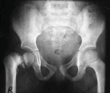
Figure 9.2
AP radiograph of a patient with idiopathic coxa vara affecting the right hip. Note the difference in aforementioned parameters between the two hips and the presence of Fairbank’s triangle
Further investigations specific to skeletal dysplasias and acquired causes are performed as required.
Prognosis
For primary coxa vara, progression is dependent on the Hilgenreiner-epiphyseal angle:
<45° defect will usually heal spontaneously and will not progress.
45–60° – may progress but needs close observation.
>60° will progress.
In acquired forms there should be minimal risk of progression provided the underlying cause is corrected.
Treatment
This is also based on Hilgenreiner-epiphyseal angle:
<45° – no treatment required – should resolve spontaneously.
45–60° – observe – consider osteotomy if severe gait abnormality or progressive.Related posts:

Stay updated, free articles. Join our Telegram channel

Full access? Get Clinical Tree


