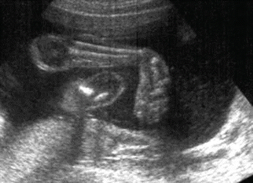
Figure 12.2
An antenatal ultrasound scan demonstrating a clubfoot
Radiographic analysis can be obtained to differentiate from a vertical talus, although X-rays are less commonly used for diagnosis and treatment currently (Fig. 12.3).
On The AP view, the following observations can be made:
1.
Lines drawn through the long axis of the calcaneum and talus will give a talo-calcaneal (Kite) angle of less than 20° (in a normal foot this would be between 20 and 40°). This is known as “ parallelism” of the talus and calcaneum.
2.
Lines are drawn down the long axis of the first metatarsal and the talus. In a foot with talipes the angle is “negative” or in adduction; in a normal foot, it is in slight abduction – 30°.
3.
There is also medial displacement of the cuboid ossification centre.
On the lateral radiograph, lines are drawn through the long axis of the talus and the inferior margin of the calcaneum – in a patient with CTEV, the angle is less than 25°.
Grading
The Pirani grading system is helpful to monitor initial treatment and early progress, and is the most commonly employed clinically (Table 12.1). It involves scoring based on six clinical signs of contracture with 0 being no abnormality, 0.5 mild abnormality and 1 severe abnormality.
The areas assessed are:
The hindfoot:
1.
Severity of the posterior crease.
2.
Emptiness of the heel pad.
3.
Rigidity of the equinus.
In the midfoot:
1.
Curvature of the lateral border of the foot.
2.
Severity of the medial crease.
3.
Position of the lateral head of the talus.
The other well known scoring system, the Dimeglio system, scores four essential parameters based on severity and four “pejorative elements”, but this system is rarely used in contemporary treatment.
Management
Although both operative and non-operative methods have been employed in the treatment of clubfoot, the mainstay of management is now based around non-operative methods, in particular the Ponseti method. It involves serial casting with or without an Achilles tenotomy, followed by the use of ‘boots and bars’. The French functional physiotherapy method also has good results, although it is a far more intensive treatment regimen.
The manoeuvres, which precede serial casting, target specific elements of the clubfoot deformity. The order in which they are performed is important, following the acronym CAVE (cavus, adductus, varus, equinus) (Fig. 12.4).
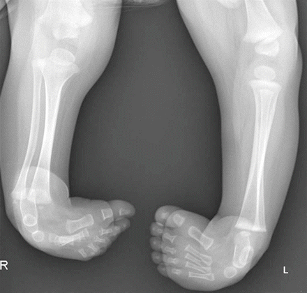
Figure 12.3
Radiograph of the lower limbs in a patient with bilateral clubfoot deformity
The first manoeuvre to correct the deformity is to align the first ray with the rest of the metatarsals in order to correct the cavus. This is followed by casting with lateral pressure on the talar head, in order to correct the forefoot adductus and hind-foot varus. At this point, an Achilles tenotomy is performed in the majority (≈90%), after which the equinus is corrected in the final cast.
Related posts:

Stay updated, free articles. Join our Telegram channel

Full access? Get Clinical Tree


