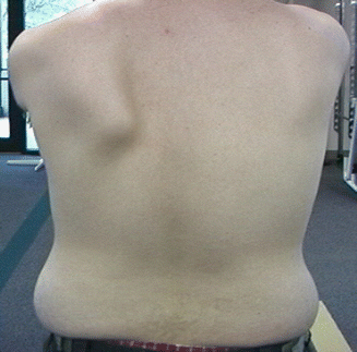
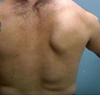
Fig. 28.1
Dynamic assessment to identify presence of scapular dyskinesis
28.6.3.3 Manual Muscle Testing
One test advocated to assess the integrity of the lower trapezius and serratus anterior muscles is that of the low row [57]. To perform this maneuver, the patient is standing with the involved arm resting at the side with the palm facing posteriorly (Fig. 28.2). The patient is instructed to extend their trunk and push their hand maximally against an examiner’s resistance in the direction of shoulder extension and instructed to retract and depress the scapula . This maneuver assesses both muscles’ ability to actively stabilize the scapula while providing the examiner a visual depiction of lower trapezius muscle contraction. Other tests such as active scapular squeezing or pinching (rhomboids and middle trapezius) and the wall push-up (serratus anterior) have also been advocated as maneuvers to employ to assess scapular muscle function.


Fig. 28.2
Low row maneuver to isometrically assess lower trapezius strength
28.6.3.4 Posture and Flexibility
Coracoid based inflexibility can be assessed by palpation of the pectoralis minor and the short head of the biceps brachii at their insertion on the coracoid tip. They will usually be tender to palpation, even if they are not symptomatic in use, can be traced to their insertions as taut bands, and will create symptoms of soreness and stiffness when the scapulae are manually maximally retracted and the arm is slightly abducted to approximately 40–50. A rough measurement of pectoralis minor tightness may be obtained by standing the patient against the wall and measuring the distance from the wall to the anterior acromial tip. This can be done using a “double square” device with a patient standing with his or her back against a wall [67]. A bilateral measurement is taken (in inches or centimeters) to determine if there is a notable difference between the involved and noninvolved shoulder, with a side to side asymmetry >3 cm considered abnormal.
To obtain accurate glenohumeral internal rotation measurements, the patient should be positioned supine on a flat level surface. A second examiner should be positioned behind the patient in order to properly stabilize the scapula by applying a posteriorly directed force to the coracoid and humeral head to ensure that scapular movement does not occur (Fig. 28.3). The humerus is supported on the surface with the elbow placed at 90° and the arm on a bolster in the plane of the scapula. A measurement is obtained using a standard bubble goniometer where the fulcrum is set at the olecranon process of the elbow, the stationary arm perpendicular to the table as documented by the bubble on the goniometer, and the moving arm in line with the styloid process of the ulna. The clinician passively moves the arm into internal and external rotation. Rotation is taken to “tightness,” a point where no more glenohumeral motion would occur unless the scapula would move or the examiner applies rotational pressure. This measurement should be taken bilaterally, and side to side differences are calculated. Side-to-side differences in internal rotation greater than 20° are considered a pathologic glenohumeral internal rotation deficit (GIRD) [68].


Fig. 28.3
Preferred patient for humeral internal rotation goniometric measurement
28.6.3.5 Symptom Alteration (Corrective Maneuvers)
If scapular dyskinesis is demonstrated on the clinical exam of patients with shoulder injury, different types of corrective maneuvers may be employed to determine the effect of the altered motion on symptoms or signs of shoulder injury. The goal of the maneuvers would be to alter or reduce some of the signs or symptoms.
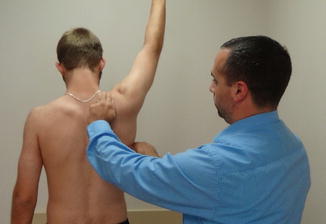
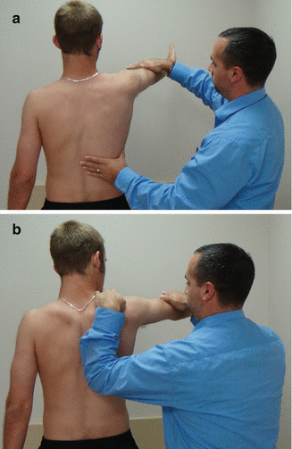
The scapular assistance test (SAT) and scapular retraction test (SRT) are corrective maneuvers that may alter the injury symptoms and provide information about the role of scapular dyskinesis in the total picture of dysfunction that accompanies shoulder injury and needs to be restored [35]. The SAT helps evaluate scapular contributions to impingement and rotator cuff strength, and the SRT evaluates contributions to rotator cuff strength and labral symptoms. In the SAT, the examiner applies gentle pressure to assist scapular upward rotation and posterior tilt as the patient elevates the arm (Fig. 28.4) [69]. A positive result occurs when the painful arc of impingement symptoms is relieved and the arc of motion is increased. This test has good test/retest reliability [70]. The test has been found to alter scapular motion by increasing scapular posterior tilt [71], so a positive test would point to the need for improvement in pectoralis flexibility and lower trapezius strength. In the SRT, the examiner grades the supraspinatus muscle strength following standard manual muscle testing procedures or by a hand held dynamometer (Fig. 28.5a). The clinician then manually places and stabilizes the scapula in a retracted position (Fig. 28.5b). A positive test occurs when the demonstrated supraspinatus strength is increased or the symptoms of internal impingement in the labral injury are relieved in the retracted position [23]. The major kinematic result of this test is to increase scapular external rotation and posterior tilt, so a positive test would indicate that rotator cuff strengthening is not necessary, and focus should be on rhomboid strengthening and serratus function in retraction. Although these tests are not capable of diagnosing a specific form of shoulder pathology, a positive SAT or SRT shows that scapular dyskinesis is directly involved in producing the symptoms and indicates the need for inclusion of early scapular rehabilitation exercises to improve scapular control.


Fig. 28.4
Scapular assistance test to demonstrate scapular involvement in shoulder dysfunction

Fig. 28.5
(a) Scapular retraction test begins by manually muscle testing forward elevation. (b) The scapula is manually stabilized in retraction and the arm is retested to determine if scapular positioning is affecting the manual muscle test result
28.7 Summary
Normal scapular mobility and stability are key and basic components of normal shoulder function. Scapular dyskinesis, alteration in mobility and stability, has been associated with clinical dysfunction in a variety of shoulder injuries. It may be a cause or an effect. Evaluation for and restoration of scapular dyskinesis can help provide information regarding treatment guidelines, demonstrate areas of emphasis in rehabilitation, and indicate progress towards functional restoration.
References
1.
Bagg SD, Forrest WJ. A biomechanical analysis of scapular rotation during arm abduction in the scapular plane. Am J Phys Med Rehabil. 1988;67:238–45.PubMed
2.
Inman VT, Saunders JB, Abbott LC. Observations of the function of the shoulder. Clin Orthop Relat Res. 1996;330:3–13.PubMed
3.
Bagg SD, Forrest WJ. Electromyographic study of the scapular rotators during arm abduction in the scapular plane. Am J Phys Med. 1986;65(3):111–24.PubMed
4.
Speer KP, Garrett WE. Muscular control of motion and stability about the pectoral girdle. In: Matsen Iii FA, Fu F, Hawkins RJ, editors. The shoulder: a balance of mobility and stability. Rosemont: American Academy of Orthopaedic Surgeons; 1994. p. 159–73.
5.
Lukasiewicz AC, McClure P, Michener L, Pratt N, Sennett B. Comparison of 3-dimensional scapular position and orientation between subjects with and without shoulder impingement. J Orthop Sports Phys Ther. 1999;29(10):574–86.PubMed
6.
McClure PW, Michener LA, Sennett BJ, Karduna AR. Direct 3-dimensional measurement of scapular kinematics during dynamic movements in vivo. J Shoulder Elbow Surg. 2001;10:269–77.PubMed
7.
Ludewig PM, Cook TM, Nawoczenski DA. Three-dimensional scapular orientation and muscle activity at selected positions of humeral elevation. J Orthop Sports Phys Ther. 1996;24(2):57–65.PubMed
8.
Ludewig PM, Behrens SA, Meyer SM, Spoden SM, Wilson LA. Three-dimensional clavicular motion during arm elevation: reliability and descriptive data. J Orthop Sports Phys Ther. 2004;34(3):140–9.PubMed
9.
Ludewig PM, Phadke V, Braman JP, Hassett DR, Cieminski CJ, LaPrade RF. Motion of the shoulder complex during multiplanar humeral elevation. J Bone Joint Surg Am. 2009;91A(2):378–89.
10.
Sahara W, Sugamoto K, Murai M, Yoshikawa H. Three-dimensional clavicular and acromioclavicular rotations during arm abduction using vertically open MRI. J Orthop Res. 2007;25:1243–9.PubMed
11.
Kibler WB, Chandler TJ, Shapiro R, Conuel M. Muscle activation in coupled scapulohumeral motions in the high performance tennis serve. Br J Sports Med. 2007;41:745–9.PubMedCentralPubMed
Stay updated, free articles. Join our Telegram channel

Full access? Get Clinical Tree


