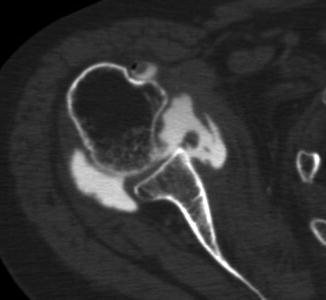
Fig. 12.1
Fast spine-echo proton density-weighted axial image. The articular cartilage and the labrum are clearly visualized without injecting Gd into the glenohumeral joint on this 3.0-T MRI . The posterior labrum attaches to the glenoid through a thick cartilage, which may mimic a posterior labral tear from the glenoid. The anterior labrum also attaches to the glenoid rim through the articular cartilage
12.3 MR Arthrography
MR arthrography clearly depicts the contrast between Gd and the intra-articular structures such as the labrum , articular cartilage, and the joint capsule (Fig. 12.2). Between the anterior capsule and the anterior labrum is the middle glenohumeral ligament, which runs parallel to the anterior labrum .
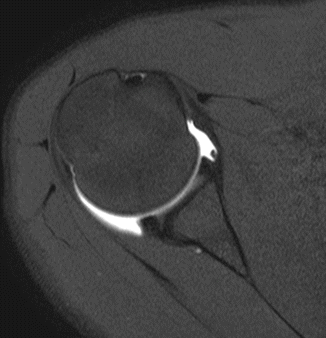

Fig. 12.2
Fast spine-echo T1-weighted axial image. The anterior and posterior labra, articular cartilage, the middle glenohumeral ligament, and the joint capsule were clearly visible with the help of the contrast material (Gd)
For the purpose of assessing the labrum , the coronal oblique and axial images have limitations. When the slices are cut perpendicular to the labrum , its attachment to the glenoid rim is most clearly visible. In order to achieve this best image for any portion of the labrum , the radial-sequence MR imaging has been introduced [15]. The scout view of the glenoid shows the orientation of the slices (Fig. 12.3). Using these slices, any portion of the labral attachment to the glenoid is clearly visualized. The slice passing through 12–6 o’clock is equivalent to a conventional coronal oblique image (Fig. 12.4). On this image, the superior labral tear is observed. On the 2–8 o’clock slice, both the anteroinferior and posterosuperior labra are detached from the glenoid (Fig. 12.5). On the 4–10 o’clock slice, the anterosuperior labrum is detached from the glenoid (Perthes lesion ) (Fig. 12.6). Other types of anterior labral lesion such as an ALPSA (anterior labroligamentous periosteal sleeve avulsion) lesion [16] (Fig. 12.7) and a GLAD (glenolabral articular disruption) lesion [17] (Fig. 12.8) are clearly depicted on these MR arthrograms. The posterior inferior part of the labrum may have a concealed tear of the labrum in cases with multidirectional or posteroinferior shoulder instability (Fig. 12.9). This concealed tear is called a Kim lesion [11]. A new imaging technique, 3-D reconstruction of MR images, shows that the SLAP lesion extends all the way down to 8 o’clock anteriorly and to 2 o’clock posteriorly (type V SLAP lesion ) (Fig. 12.10).
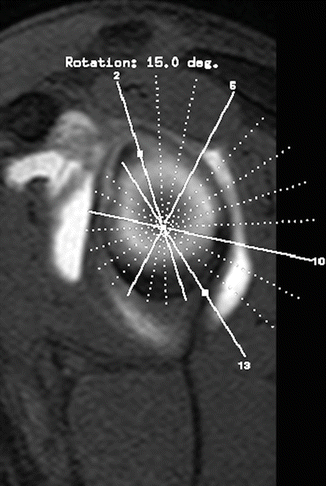
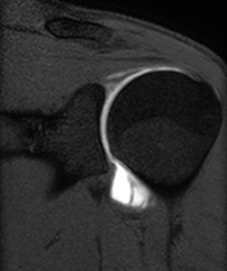
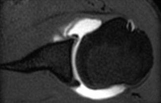
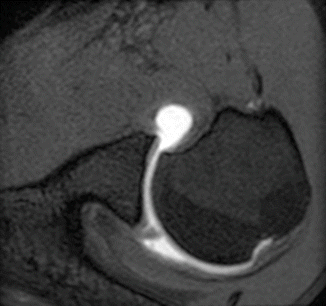
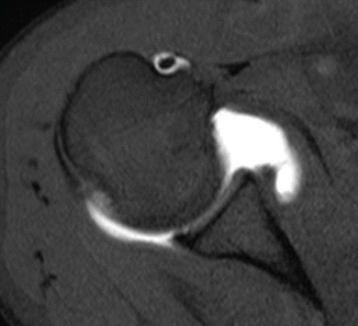
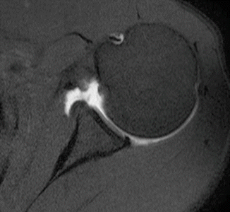
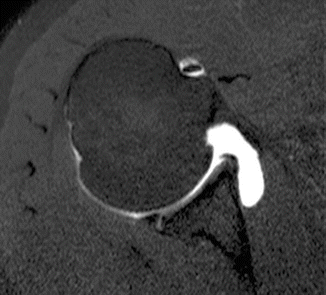
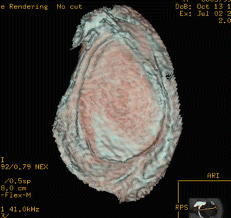

Fig. 12.3
Scout view showing the orientation of radial slices. The slices are drawn through the center of the glenoid with a 15-degree increment. The slice passing through 12–6 o’clock is equivalent to a conventional coronal oblique image, and the slice passing through 3–9 o’clock is equivalent to a conventional axial image

Fig. 12.4
Fast spine-echo T1-weighted 12–6 o’clock slice. There is a tear of the superior labrum at 12 o’clock position (left shoulder)

Fig. 12.5
Fast spine-echo T1-weighted 2–8 o’clock slice. The anteroinferior labrum is detached from the glenoid at 8 o’clock position, and the posterosuperior labrum is also detached at 2 o’clock (left shoulder)

Fig. 12.6
Fast spine-echo T1-weighted 4–10 o’clock slice. There is a tear of the anterosuperior labrum at 10 o’clock position (left shoulder)

Fig. 12.7
ALPSA (anterior labroligamentous periosteal sleeve avulsion) lesion. The anterior labrum together with the anterior capsule including the inferior glenohumeral ligament is displaced medially. This is called an ALPSA lesion , which causes anterior instability of the shoulder

Fig. 12.8
GLAD (glenolabral articular disruption) lesion. The anterior labrum is detached and displaced medially with associated articular cartilage damage

Fig. 12.9
Kim lesion. The posterior inferior part of the labrum may have a concealed tear of the labrum in cases with multidirectional or posteroinferior shoulder instability. This concealed tear is called a Kim lesion

Fig. 12.10
3D reconstruction of an en face view of the glenoid and labrum. This is the left shoulder with 9 o’clock being anterior and 3 o’clock being posterior. The SLAP lesion extends all the way down to 8 o’clock anteriorly and to 2 o’clock posteriorly (type V SLAP lesion)
12.4 CT Arthrography
In order to make clear contrast among the soft tissues, CT arthrography is performed with use of contrast medium such as iodine contrast, air, or both (double contrast CT arthrography). This CT arthrogram with iodine contrast reveals that the anterior labrum separated from the anterior capsule and from the glenoid rim (Bankart lesion ) (Fig. 12.11). The anterior articular cartilage looks thinner than the posterior one due to recurrent anterior dislocations of the shoulder. When both the iodine contrast and the air are used as contrast materials, it is called double-contrast CT arthrography (Fig. 12.12), which shows the surface of the intra-articular structures very clearly. This is also a case of recurrent anterior dislocation, with the anterior labrum and a part of the inferior glenohumeral ligament disappearing (Bankart lesion). The articular cartilage looks intact.
