Fig. 23.1
Fast spin-echo T2-weighted oblique coronal image 1. This anterior slice depicts the subscapularis tendon with its several intramuscular tendons spanning into the muscle belly. Above the subscapularis tendon is the coracoid process with the conjoint tendon running vertically

Fig. 23.2
Fast spin-echo T2-weighted oblique coronal image 2. This middle slice depicts the anterior portion of the supraspinatus tendon with a thick single intramuscular tendon (dark band). It attaches to the superior facet of the greater tuberosity, which looks horizontal on this image. There is a small amount of fluid in the glenohumeral joint, acromioclavicular joint, and the subacromial bursa. The mid portion of the glenoid is also depicted on this image
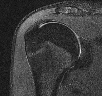
Fig. 23.3
Fast spin-echo T2-weighted oblique coronal image 3. This middle posterior image depicts the superior portion of the infraspinatus tendon that attaches to the middle facet of the greater tuberosity, which looks slightly inclined on this image. The intramuscular tendon (dark band) is observed on the articular side of the tendon. The posterior portion of the glenoid is also depicted on this image
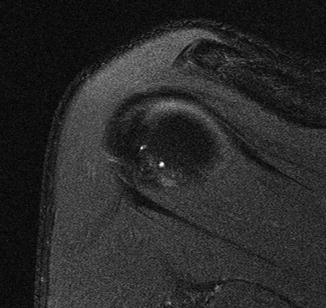
Fig. 23.4
Fast spin-echo T2-weighted oblique coronal image 4. This posterior slice shows the posterior portion of the humeral head with the superior portion of the infraspinatus tendon superiorly and the teres minor tendon inferiorly. There are two small bony cysts (high signal) on the posterior aspect of the humeral head
The most lateral slice of the sagittal oblique view shows the greater tuberosity with distal cross section of the supraspinatus tendon superiorly, infraspinatus tendon posterosuperiorly, and teres minor tendon posteriorly (Fig. 23.5). More medially, the lesser tuberosity with the cross section of the subscapularis tendon is observed (Fig. 23.6). On this image, the muscle bellies of the supraspinatus, infraspinatus, and teres minor are observed with their intramuscular tendons as dark signal areas inside the muscle belly. The medial slice at the level of the glenoid surface depicts the cross sections of all the rotator cuff muscles with their intramuscular tendons located in the middle of the muscle bellies (Fig. 23.7).
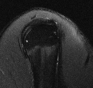
Fig. 23.5


Fast spin-echo T2-weighted oblique sagittal image 1. This lateral slice shows the sagittal view of the greater tuberosity with distal cross sections of the supraspinatus tendon superiorly, the infraspinatus tendon posterosuperiorly, and the teres minor tendon posteriorly. Anteriorly there runs the tendon of the long head of the biceps vertically along the bicipital groove. A small bony cyst is depicted close to the surface of the posterior aspect of the humeral head
Related posts:


Stay updated, free articles. Join our Telegram channel
Full access? Get Clinical Tree
