Fig. 9.1
Left cadaveric shoulder demonstrating the intra-articular ligaments
The coracohumeral ligament originates proximally at the dorsolateral aspect of the coracoid as a fibrous brand extending superiorly over the head of the humerus before it attaches to the greater tuberosity [5, 37]. This ligament then blends in with the superior glenohumeral ligament (SGHL) inferiorly. The SGHL (arthroscopic view, Figs. 9.2 and 9.3) originates just anterior to the insertion of the long head of the biceps at the superior portion of the glenoid. The SGHL is described as originating between the 12 and 2 o’clock positions on the glenoid, just anterior to the insertion of the long head of the biceps, and runs inferiorly and laterally where it then inserts into the humerus at the fovea capitis which lies slightly superior to the lesser tuberosity [8, 45]. As we move counterclockwise around the glenoid, the middle glenohumeral ligament (MGHL, arthroscopic view Fig. 9.4) originates just inferior to the SGHL [5, 8]. It then inserts medial to the lesser tuberosity on the humerus. The MGHL often follows the superior marginal of the subscapularis tendon; however, it has also been described as the most variable ligament of the labral structures of the shoulder [5, 8]. Anatomically, the MGHL may appear to be flush to the SGHL, as a distinct structure that arises from the inferior aspect of the SGHL as described above, or may not even be clearly identified. The inferior segment of the capsule, the inferior glenohumeral ligament has been identified as the broad inferior glenohumeral ligament and is further broken down into an anterior band which originates from the 3 to 5 o’clock position, posterior band which originates from the 7 to 9 o’clock position which inserts onto the posterior articular margin of the humerus, and the axillary pouch in between [5, 8, 35].
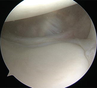
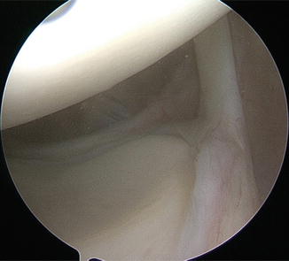
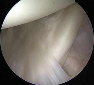

Fig. 9.2
Arthroscopic image of anterior-superior and anterior-inferior labrum with anterior capsule structures. Patient is in the lateral position, image taken from the posterior portal

Fig. 9.3
Arthroscopic image of the superior biceps labral complex showing posterior and anterior superior labrum . Patient is in the lateral position, image taken from the posterior portal

Fig. 9.4
Arthroscopic image of the middle glenohumeral ligament coming off at 90° angle to the subscapularis tendon. Patient is in the lateral position, image taken from the posterior portal
The labrum of the glenoid circumferentially surrounds the outer portion of the glenoid and is the point of insertion of all the glenohumeral ligamentous structures described above. It acts to deepen the socket on average 9 mm in the superior-inferior dimension and 5 mm in the anteroposterior plane which acts to add stability to the joint [35]. The only caveat to these insertions is in regard to the IGHL which has two insertion sites. One insertion is into the anteroinferior aspect of the labrum with a second insertion into the anterior neck of the glenoid [8]. An additional structure that inserts into the superior aspect of the glenoid, just superior to the SGHL, is the long head of the biceps tendon . The biceps tendon originates distally at the radial tuberosity. It course up the humeral shaft in the bicipital groove located between the great and lesser tuberosities. The tendon finally inserts into the superior glenoid and is held in place by the coracohumeral ligament superiorly (arthroscopic view, Fig. 9.5) and the superior glenohumeral ligament inferiorly [46, 48]. This anatomic description has been termed the biceps pulley for the long head of the biceps.
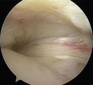

Fig. 9.5
Arthroscopic image of the biceps sling. Patient is in the lateral position, image taken with 70° scope in the posterior portal
The glenoid is a pear-shaped shallow socket with a wider inferior half [35, 37]. This structure is tilted anteriorly therefore making the joint more susceptible to anterior dislocation [37]. Specifically, the physical features of the glenoid bone are often variable but have been reported as 39 mm (±3.7 mm; range 30–48 mm) in the superior-inferior dimension, 29 mm (±3.1 mm; range 21–35 mm) in the anteroposterior dimension of the lower half, and 23 mm (±2.7 mm; range 18–30 mm) in the anteroposterior dimension of the upper half with the fossa slightly inclined and retroverted [8, 35]. The variation of the size and angle of the glenoid is part of what makes pathology of this bone challenging to treat.
9.3 Histological Anatomy of the Labrum
Histology has provided a greater insight into the anatomic detail of the glenoid and labrum. At the 10–12 o’clock position, the labrum is positioned off the rim of the glenoid and has a concave surface to articulate with the humeral head [2]. There is also a synovial-lined recess between the labrum and the supraglenoid tubercle (approximately 6 mm off glenoid face), allowing the superior labrum to be relatively mobile (Fig. 9.6). The stability of the superior labrum and the glenohumeral joint is further enhanced by the biceps tendon (dynamic stability) and SGHL (static stability) [2, 12].
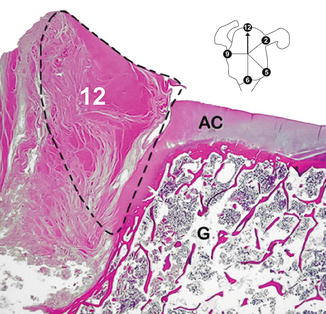

Fig. 9.6
Histological axial section at 12 o’clock. AC articular cartilage, G glenoid cancellous bone, L labrum . Note the labral attachment is off the face of the glenoid
In contrast, the inferior labrum overlaps the glenoid face by 4 mm, with a convex rounded cross section which increases the depth by up to 50 % ([2, 18, 24] (Figs. 9.7 and 9.8). The inferior labrum is adherent to the articular cartilage and has a rigid bony foundation which prevents mobility of the labrum. The inferior labrum provides a bumper effect and is a fixed organ designed for compression. This anterior inferior labrum is critical to stability and is the common site of the Bankart labral tear seen in traumatic instability (Fig. 9.9).
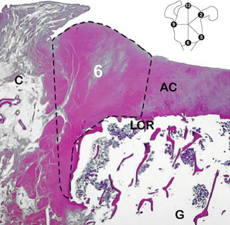
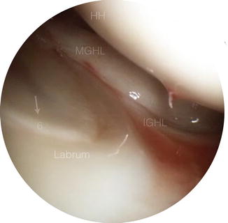
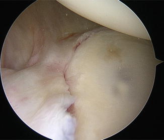

Fig. 9.7
Histological axial section at 6 o’clock. AC articular cartilage, C capsule, G glenoid cancellous bone, L labrum , LCR internal labral circumferential ridge. Note the bumper effect of the labrum , which is firmly attached on the face of the glenoid

Fig. 9.8
Arthroscopic image of intact inferior labrum. HH humeral head, MGHL medial glenohumeral ligament, IGHL inferior glenohumeral ligament, and “6” correlating to the 6 o’clock position of the labrum

Fig. 9.9
Arthroscopic image of the torn anterior band of the inferior glenohumeral ligament. Patient is in the lateral position, image taken from the superior portal
There are three layers in the labrum: a thin mesh-like superficial layer, a stratified middle layer, and a circumferential deep layer [29]. The fibers that merge with the articular cartilage and capsule are radially orientated [2]. The radial collagen fibers at the superior labrum–articular interface are less densely packed allowing greater mobility of the labrum. In addition to the dense collagen fibers, there are chondrocytes and elastic fibers [24, 26, 27, 29]. With aging, there is a loss of chondrocytes [34].
The labrum and bone interface is similar to a tendon insertion with a narrow zone of transition through uncalcified fibrocartilage, to calcified fibrocartilage incorporating Sharpey’s fibers, to bone (Fig. 9.10). The labrum–capsule interface merges imperceptibly, which explains why the labral tears often occur between the labrum and bone. They are both composed of fibrous tissue with the labrum being much denser.
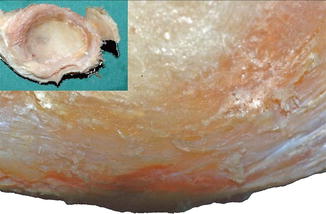

Fig. 9.10
The labrum has been stripped from the glenoid face to demonstrate circumferential striations from the Sharpey’s fiber insertion
The biceps tendon fibers pass directly through the labrum to the bony insertion via the shortest possible route. The biceps tendon inserts both into the supraglenoid tubercle over a wide area (6 mm coronal section) and to the superior labrum. Approximately two thirds of the fibers insert into the supraglenoid tubercle and the remaining fibers attach to the superior labrum.
9.4 Functional Anatomy
The stability of the glenohumeral joint relies on a combination of muscles, bones, and other structures to maintain its integrity. Passive stability to the joint is provided by the labrum , capsule, and glenohumeral ligaments in normal shoulder function [11, 40]. The glenoid labrum is a fibrocartilaginous structure that encircles the edge of the glenoid fossa and acts to simultaneously protect the outer edges of the glenoid while also deepening the pocket to protect against dislocation [25–27, 32]. This labrum is continuous with the capsule, a thin structure that surrounds the joint by attaching to the glenoid medially and around the anatomic neck of the humerus laterally. The capsule allows for extensive range of motion. By itself, the thin, loose-fitting structure of the capsule does not offer much stability; however, the capsule is reinforced by the glenohumeral ligaments, which provide critical static restraints to dislocation [11, 40]. There are three glenohumeral ligaments that run within the capsule to provide support, all of which were described in the physical anatomy section. The functional significance of these structures will be described in detail within this section.
The superior glenohumeral ligament prevents inferior translation of the adducted shoulder and is an important restraint up to 50° abduction and during external rotation [5, 47]. The middle glenohumeral ligament limits lateral rotation from 45° to 75° abduction as well as supports the joint anteriorly [49]. The inferior glenohumeral ligament complex, which is composed of the anterior band, axillary pouch, and posterior band, is a vital stabilizer for the glenohumeral joint [30]. The anterior band is responsible for restraining anterior and inferior translation of the humerus [41, 45]. Support by the anterior band is most effective at increased (greater than 75°) abduction. This structure plays an important role in sports by preventing anterior humeral head migration when the arm is abducted to 90° and externally rotated. This is evident in activities such as freestyle stroke, tennis serve motion, and any overhead arm position [30]. Alternatively, the posterior band provides posterior stability primarily when the arm is in flexion and internal rotation [5].
Stay updated, free articles. Join our Telegram channel

Full access? Get Clinical Tree


