Fig. 10.1
Arthroscopic view of the shoulder ligaments: (a) SGHL within the rotator interval. (b) MGHL as it crosses the subscapularis tendon. (c) AIGHL coursing inferiorly and obliquely
More recently, further anatomic research has led to an appreciation of the variations in these main ligaments and identification of further important ligaments. This chapter focuses on these new findings, and their clinical relevance.
10.2 Glenohumeral Ligaments
10.2.1 Incidence of Glenohumeral Ligaments
10.2.2 Variable Origins of the Glenohumeral Ligaments
The SGHL usually originates from the supraglenoid tubercle just anterior to the long head of biceps attachment. However, it has also been found to originate from the posterior aspect of the supraglenoid tubercle, the superior labrum , the long head of biceps, the MGHL or a combination of these structures [7]. The SGHL origin has been classified into three types, the most common of which was from the MGHL , long head of biceps tendon and superior labrum [8]. (Table 10.2) spans from the anterior superior glenoid to the lesser tuberosity.
Types of SGHL origin | Location | Incidence (%) |
|---|---|---|
A | MGHL , biceps tendon, superior labrum | 76 |
B | Biceps tendon, superior labrum | 21 |
C | Biceps tendon | 1 |
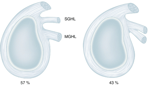
Two common variations of the MGHL origin have been reported. The first is from the labrum and SGHL (43 %) and the second from the labrum alone, independent of the SGHL (57 %) [9–11] (Fig. 10.2).


Fig. 10.2
Common variations in the origin of the MGHL
The AIGHL spans from the anterior glenoid and labrum to the lesser tuberosity and the surgical neck of the humerus [1, 2]. The AIGHL most commonly originates from the 3 o’clock position of the glenoid but can be as proximal as 2 o’clock and as distal as 5 o’clock (Table 10.3) [11–14].
The PIGHL usually arises from the posterior aspect of the glenoid between 7 and 9 o’clock. It is more difficult to appreciate than the AIGHL and is confluent with the thinner posterior capsular structures [14].
10.2.3 Variations of the Middle Glenohumeral Ligament
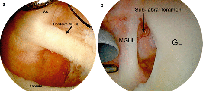
The MGHL arises from the upper anterior aspect of the glenoid, runs obliquely and deep to the subscapularis tendon to insert onto the lesser tuberosity. A number of variants have been described including the sublabral foramen (12 %), cord-like MGHL (18 %) and the Buford complex (1–2 %) [1, 2, 9, 15, 16]. With a cord-like MGHL , the medial and lateral edges of the MGHL tend to have a curling shape. The Buford complex (Fig. 10.3a, b) is defined as an absent anterosuperior labrum with a cord-like MGHL , which originates from the superior labrum, and inserts into the humerus. The anterior labrum superior to the glenoid equator is variable in the degree of its attachment and the presence of a sublabral foramen is observed in around 12 % of arthroscopies [17]. This is a localised area where there is no labral attachment to the glenoid and should not be confused with a pathologic lesion such as a Bankart tear which has an irregular edge, associated synovitis, capsular tears and extends more inferiorly [15, 17].


Fig. 10.3
(a) A cord like MGHL present in a Buford complex. (b) An anterior superior sublabral foramen. SS Subscapularis tendon, GL Glenoid (Copyright Yin, OCNA [17])
10.2.4 Variations of the Inferior Glenohumeral Ligament
O’Brien et al. described three components of the IGHL : the anterior band (AIGHL), the posterior band (PIGHL), and the axillary pouch (AxIGHL), which is the hammock formed between the two bands.
Itoigawa et al. [12] reported that the mean length of the AIGHL attachment was 11.7 mm. The mean depth was 4.7 mm (1.6 mm on the articular cartilage and 3.0 mm on the glenoid neck) at the 2 o’clock position, 6.7 mm (2.4 and 4.3 mm, respectively) at the 3 o’clock position, 8.4 mm (3.0 and 5.4 mm, respectively) at the 4 o’clock position, and 6.8 mm (2.5 and 4.3 mm, respectively) at the 5 o’clock position.
Histology demonstrates that the AIGHL usually attaches to both the cartilage and bone (88 %), but can be attached to only the bone (12 %).
10.3 Rotator Interval and Its Contents
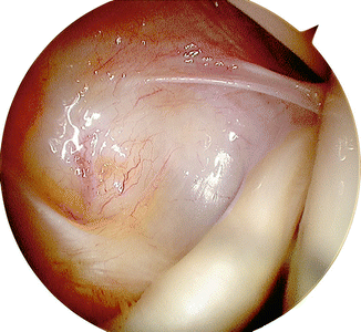
The rotator interval (RI) is a triangular space formed by the upper border of subscapularis inferiorly, the anterior edge of the supraspinatus superiorly and the coracoid process medially (Fig. 10.4) [19].


Fig. 10.4


Arthroscopic view of the rotator interval
Stay updated, free articles. Join our Telegram channel

Full access? Get Clinical Tree


