Femoral diaphyseal fractures
Epidemiology of femoral fractures in the elderly
Classification and mechanism of injury
Assessment and preparation for surgery
Intramedullary nailing in elderly femoral shaft fractures
Plating in elderly femoral shaft fractures
INTRODUCTION
The femur is the longest and strongest bone in the human body. Fractures of the shaft of the femur are usually thought to be due to high velocity injuries, primarily after motor vehicle accidents or after falls from a height. There is increasing awareness that in the elderly these fractures are of an osteoporotic nature.1,2 As opposed to fractures in younger groups, they typically involve females above the age of 70, usually with minimal trauma. This group of people requires the same detailed workup as any other osteoporotic fracture.
There is also a small subset of fractures in the elderly that are due to malignancy and metabolic bone diseases as well as stress fractures. These, although rare, must be considered in all cases despite innocuous-looking X-rays. Where appropriate, additional blood tests and imaging must be ordered.
There is a paucity of well-conducted studies in the elderly femoral shaft fracture. What little evidence we have suggests that they have approximately the same mortality and morbidity as a hip fracture in a patient of the same age. With a rising age distribution in most developed countries, these fractures are likely to increase in incidence as well as absolute numbers.
The vast majority of these fractures are treated surgically except in the very medically unfit. In general they should be treated along similar principles as hip fractures with the aim of allowing early weight bearing and rehabilitation.
EPIDEMIOLOGY OF FEMORAL FRACTURES IN THE ELDERLY
Femoral fractures exhibit a bimodal distribution. This epidemiological pattern has been demonstrated by numerous authors including Singer and Hedlund.3,4 While the average incidence has been estimated at 1–1.33 fractures per 10,000 population per year, Singer has shown that cases clustered around the 15–34-year-old age group [incidence of 1.64–3.73 per 10,000 population] and started to peak again after 70 years of age [incidence of 2.3–37.14 per 10,000 population] in femoral shaft fractures presenting to the Royal Infirmary of Edinburgh from 1992 to 1993.3 Chapter 1 in this book shows that currently 69.9% of all patients who present to the Royal Infirmary of Edinburgh with femoral diaphyseal fractures are ≥65 years of age with 84% of females being ≥65 years of age.
In addition to the bimodal distribution, a gender specific pattern of presentation has also been demonstrated by Singer3 and Hedlund.4 Singer’s younger cohort clustered in male patients while the older cohort involved a far higher proportion of female patients.3 In another Swedish cohort spanning 1998–2004, men had a younger median age (27 years, IQR 12–68),5 whereas women had a far higher median age (79 years, IQR 62–86),5 similar to those sustaining osteoporotic hip fractures. Analysis showed that 54% of the admissions were females and 46% males in this cohort.5
Although much attention has been focused on the epidemiological trends, prevention and management of proximal femoral fractures, diaphyseal femoral fractures in the elderly may carry an equivalent impact. Comparing a cohort in the 1950s to one in the 1970s and early 1980s, Bengnér and co-authors noted that the risk of low energy femoral shaft fractures had increased in elderly women.6 These patients may be more frail and require more healthcare resources as evidenced by up to 85% of patients presenting with low energy femoral fractures having multiple comorbidities, with a length of hospital stay (15 days) equivalent or even longer than that of osteoporotic hip fracture cohorts. With a rapidly aging population in most developed countries, we are likely to see an increasing trend in the presentation of elderly diaphyseal femoral fractures which will place a strain on healthcare systems.
A decline in hip fracture incidence (600/100,000 person-years to 400/100,000 person-years) has been observed from 1996 to 2006 in national discharge and medical claims data in the United States, possibly as a result of aggressive preventive measures for osteoporotic fractures. In contrast, subtrochanteric, femoral shaft and lower femoral fracture rates remained stable, although at far lower rates of 20 per 100,000 person-years. Similar trends but lower rates were observed in males than females.2
CLASSIFICATION AND MECHANISM OF INJURY
There is no specific classification for elderly femoral diaphyseal fractures. The AO/OTA classification still remains the most common classification system used to categorize these fractures. In the AO/OTA classification, type A fractures are simple fractures and include spiral fractures (A1), oblique fractures (A2) and transverse fractures (A3). Type B fractures are wedge fractures and include spiral wedges (B1), bending wedges (B2) and fragmented wedges (B3). Type C fractures are complex fractures with the C1 group containing all spiral fractures, the C2 group all segmental fractures and the C3 group all comminuted fractures. In type A and B fractures the suffix 0.1 represents a fracture in the subtrochanteric zone, with 0.2 used for the middle zone and 0.3 for the distal zone. In type C fractures the suffixes 0.1 though 0.3 represent increasing bone damage.
It is important to distinguish high energy osteoporotic fractures from low energy fractures that happen to occur in the elderly. A small percentage of femoral diaphyseal fractures in the elderly result from polytrauma, usually as a result of motor vehicle accidents. These elderly patients sustaining a femoral fracture in a high energy injury behave like younger patients with a similar injury except that they have less physiological reserves. They also may have a more prolonged rehabilitation period and more trouble coping with rehabilitation.
Low energy osteoporotic femoral diaphyseal fractures
An AO/OTA A1 spiral fracture involving the middle third of the femoral shaft was reported as the most common pattern in low energy femoral shaft fractures in the mid-1990s (Figure 36.1a). These fractures were closed with no or minimal comminution.7 Such fractures are believed to occur as a result of a twisting force in osteopenic bone. Two-thirds of the patients had at least one local or general factor weakening the mechanical strength of the bone. In the majority of these patients, the femoral fracture is an isolated injury with no associated injuries.7
Age-related bone loss together with weakening of bone stock and quality from associated comorbid conditions contribute to the majority of these fractures. Aging also goes hand-in-hand with other pathophysiological changes and conditions that can predispose to femoral shaft fractures. They can be broadly classified into stress fractures arising from structural or biochemical abnormalities, pathological fractures from metastatic or primary bone diseases, metabolic disorders affecting the bone and periprosthetic fractures.
Atypical femoral fractures
An interesting shift in the pattern of femoral diaphyseal fractures in the elderly began to emerge in the mid-2000s. In contrast to the spiral pattern previously reported, these fractures had a transverse or short oblique configuration.8 (AO/OTA A3 configuration), with characteristic beaking and a medial spike of varying length and hardly any comminution (Figure 36.1b). These almost pathognomonic features have formed the basis of the American Society for Bone and Mineral Research (ASBMR) Task Force criteria for the definition of atypical femoral fractures (AFFs), a term implying deviation from the usual characteristics of the typical spiral or oblique osteoporotic femoral shaft fracture.
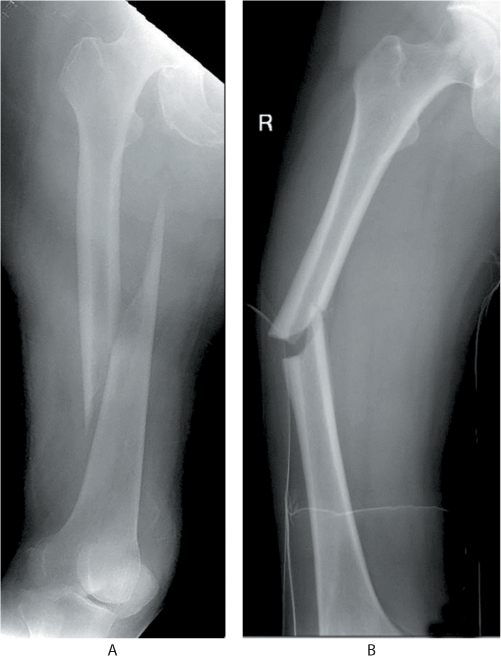
Figure 36.1 (a) Osteoporotic fracture with spiral pattern. (b) An atypical femoral fracture (AFF).
Unlike the usual osteoporotic fractures which are usually seen in the middle third but can occur anywhere along the femoral shaft, AFFs are clustered around the subtrochanteric region8 and the femoral shaft.8 They are rarely seen beyond the middle third of the shaft8 and they almost exclusively involve the tensile stress regions of the femur. These fractures usually occur as a result of low energy falls but some are actually atraumatic. Approximately 30–50% are bilateral8 They are generally believed to originate from a lateral cortical stress fracture which manifests as localized cortical thickening,8 and the presence of a ‘dreaded black line’ across the area of thickening in association with prodromal thigh pain has been shown to be associated with a high risk of complete fracture.9 An initial lack of awareness of this condition among clinicians has led to the misdiagnosis of spinal stenosis or arthrosis of the hip or knee with referred pain even with radiological evidence of the stress lesion. Often these patients are thought to have osteoarthritis of the knee and a total knee replacement done for the wrong reasons (Figure 36.2).
These unusual features, in the presence of a known history of prolonged bisphosphonate therapy8 have caused surgeons to postulates that the AFF is a stress fracture arising from oversuppression of bone turnover by bisphosphonate therapy.
Femoral stress fractures in the elderly
Stress fractures may occur as a result of physiological bowing and severe varus secondary to end-stage arthrosis. Age-related changes in femoral morphology, in conjunction with stiffness from knee arthrosis, can result in stress fractures along the femoral shaft (Figure 36.3). A resultant medialization of body weight transfer due to femoral bowing10 and increasing knee varus can lead to tensile failures along the lateral cortex of the femoral shaft.
Pathological fractures
Pathological fractures can occur due to metastatic disease from a distant tumour or from a primary tumour arising from the bone, the most common being multiple myeloma.
METASTATIC DISEASE
Pathological fractures from metastatic bone disease, though uncommon, are encountered with increasing frequency in the young-old (reported median age of 63 years) due to improving survivorship in cancer patients. The skeleton is the third most frequent location for metastases, and cancers arising from the breast, prostate, lungs, thyroid and kidneys are known to commonly metastasize to bone, with breast cancer being the most common primary tumour. The femur is the most common long bone to be affected by bony metastasis (44%)11 with the upper third involved in 50% of cases. These fractures are consistently missed in a small number of patients and where there is an index of suspicion, additional imaging and investigations should be ordered as necessary. A large majority of these fractures are treated by closed nailing and often no biopsy is taken.
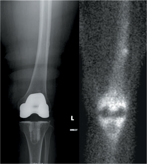
Figure 36.2 An atypical femoral fracture diagnosed as osteoarthritis of the knee with a total joint replacement done. The bone scan shows a typically hot spot on the lateral cortex.
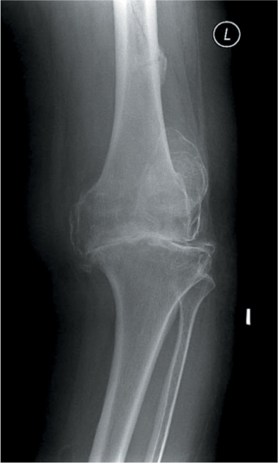
Figure 36.3 Stress fracture of the femur resulting from osteoarthritis with resulting varus and stiffness of the knee.
An impending pathological fracture of the femur is an indicator for prophylactic stabilization. The Mirel scoring system gives an estimate of fracture risk based on four parameters: the site and size of the lesion, the type of lesion and the degree of pain. Metastatic fractures are discussed further in Chapter 16.
MYELOMA
Myloma has become increasingly common in this age group and is often missed due to a low index of suspicion leading to delayed treatment and further morbidity from subsequent fractures. As many good treatment options are currently available, failure to make an early diagnosis can severely impact the patient’s long-term outcome.
METABOLIC BONE DISEASE
Many elderly patients have associated comorbidities that can result in metabolic bone disease. Common conditions include end-stage renal disease, Paget’s disease (Figure 36.4), vitamin D deficiency, malnutrition and hypoparathyroidism. An endocrinological consultation may be warranted in suspicious cases.
Periprosthetic and peri-implant fractures
The increasing incidence of arthroplasty procedures for degenerative hip and knee conditions is posing unique challenges with fractures occurring around the implants. Reported incidences of 1.1% after primary hip arthroplasty and 4.0% after revision arthroplasty, based on the Mayo Clinic Joint Registry,12 have been recorded. The average age is 68.1 years with a male:female ratio of 1:2. Interactions between the native bone and implant may influence the fracture pattern and interfere with healing or the placement of other fixation devices, and the long-term presence of the device may even change the structure of the bone and increase risk of fracture. Duncan and Masri developed the Vancouver classification according to location, implant stability and degree of bone loss to account for this complex interplay of factors and provide an algorithm facilitating treatment of these fractures. Periprosthetic fractures are discussed in Chapter 17.
Polytrauma in the elderly
As life expectancy increases in developed nations, trauma centres are projected to see an increasing load of elderly patients sustaining femoral fractures as a result of polytrauma. In cohorts matched for sex, age, Injury Severity Score (ISS) and comorbidities, the presence of a femoral fracture led to an increased number of complications, longer total hospital length of stay, more discharges to rehabilitation centres, more accompanying long bone fractures and an increased likelihood of surgery. However, there was no difference in length of ICU stay or in-hospital, 6-month and 1-year mortality between patients with and without femoral fractures.13 Polytrauma in the elderly is discussed in Chapter 14.
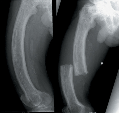
Figure 36.4 An 80-year-old patient with Paget’s disease with subsequent bowing and a ‘chalkstick’ fracture.
ASSOCIATED INJURIES
Femoral neck fractures are associated with femoral shaft fractures in 1–9% of cases. Up to 15–50%14 of these ipsilateral fractures may be missed unless specifically looked for. Femoral fractures can also have extensions to the supracondylar or intra-articular region of the distal femoral, thus limiting common treatment options such as the intramedullary nail. Careful scrutiny of radiographs of the entire femur prevent any unnecessary perioperative surprises!
Low energy femoral fractures tend to occur in isolation. An exception is seen in AFFs where contralateral involvement may result in bilateral femoral shaft fractures. A full radiographic assessment of the contralateral femur is advocated once an AFF is diagnosed.
DIAGNOSIS
The diagnosis of a fracture of the shaft of the femur is usually relatively straightforward. A plain X-ray will reveal the type and pattern of these fractures in the large majority of these fractures. However, it is vital to obtain high quality films. In large people, the standard X-ray plates may not be able to cover the ends of the femur or one end may be over-penetrated or under-penetrated. In these cases, the hip and/or knee should have a separate well centred X-ray. Both the hip and knee should be scrutinized for associated fractures.
As most osteopenic fractures in the elderly are caused by a twisting force, many of these cases result in a spiral fracture pattern that extends distally. The distal extension is often missed if not looked for and may result in suboptimal fracture fixation with a plate or nail that is too short (Figure 36.5). If in doubt, additional imaging including CT scans and/or MRI studies should be obtained.
ASSESSMENT AND PREPARATION FOR SURGERY
This group of elderly patients should be rapidly worked up and surgery performed expeditiously. Most studies show that morbidity and mortality parallel those of hip fractures.15 We recommend a similar workup, if possible in conjunction with a geriatrician and an anaesthetist. All patients should be treated as any patient with a femoral shaft fracture, with routine arterial blood gas and/or pulse oximetry monitoring, especially in the first 24 hours.
Elderly patients have a lower tolerance for blood loss and should receive crystalloid and colloid volume replacement and blood transfusions earlier than younger patients. All elderly patients should receive limb and chest physiotherapy upon admission to reduce respiratory complications and bedsores. We recommend a limb immobilizer or simple traction for pain relief if the delay until surgery is short. In cases where the time to surgical treatment is prolonged, we recommend a balanced form of traction using a skeletal traction pin inserted through the distal femur. The recommended traction weight is 15% of body weight.
TREATMENT
The aim of surgery is to restore the patient to walking and a normal lifestyle as soon as possible. Very few patients are treated non-operatively, the exception sometimes being very sick patients with a high anaesthetic risk. However we find that even bedridden patients benefit from surgery as it makes nursing, transfer and activities of daily living much easier to perform. For this reason, nailing is the treatment of choice in femoral diaphyseal fractures in the elderly. The majority of patients will undergo a closed intramedullary nailing procedure as soon as they are fit on the next operating list. We recommend against operating in the middle of the night on an emergency basis unless the patient has been fully worked up and an experienced operating team is available. Where possible, the surgical procedure should be done within 24 hours.16
Simultaneous hip and shaft fractures
Associated hip fractures are treated differently than in younger patient where priority is towards preservation of blood supply of the femoral head. In elderly patients, hip fractures, especially unstable ones, do not necessarily receive priority in treatment. A hip replacement may be done for the hip and the femoral fracture treated independently. In some cases femoral plating is warranted. If this is the case we recommend the use of locked plates.
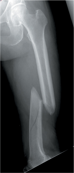
Figure 36.5 Femoral shaft fracture in an elderly patient with minimally displaced spiral extension.
Stay updated, free articles. Join our Telegram channel

Full access? Get Clinical Tree


