Fig. 2.1
Stage 15 (8.5 mm CRL) embryo transversally sectioned. Hematoxylin-Eosin stained. a (2×) and b (4×) area corresponding to the left upper limb magnified. aa axilary artery, ad anterior division of the brachial plexus, ba brachial artery, bp brachial plexus, pd posterior division of the brachial plexus, h humerus
The neural plate has divided into two divisions, anterior and posterior. The anterior one forms the musculocutaneous, ulnar and median nerves while the posterior one forms the radial nerve. It is clear that nerve ingrowth at the base of the bud begins in stage 16, but it is still not fully understood how specific nerves are guided to supply specific muscles.
The first clavicle precursor appears at 11 mm, a condensed curved connective-tissue rod stretching from acromion inwards towards the first rib [6]. The mesenchymal tissue of the humerus has now begun to chondrify, and the ulna and radius are still represented by condensed mesenchymal tissue.
Stage 17 (11–14 mm; 41 days), Stage 18 (13–17 mm; 44 days) (Fig. 2.2). The nerves are easily recognizable as far as the hand. The mesenchyme of the humerus is in a chondrified state. Haines [1947] reported that the cartilages of the shoulder and elbow joints were adopting their characteristic shapes and their trilaminar interzones at 12 mm CRL [7].
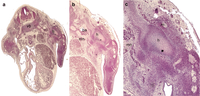

Fig. 2.2
Stage 17 (12 mm CRL) embryo transversally sectioned. Hematoxylin-Eosin stained. a (×2), b (×4) left upper limb magnified, c (×10) left shoulder magnified. ba brachial artery, mn median nerve, h humerus, rn radial nerve, vb vertebral body
The clavicle is still developing; an inner and an outer mass of precartilage is developed in the connective-tissue rod, the inner overriding above and in front the outer mass. The outer segment is at this time distinctly connected with the base of the coracoid process by the coracoclavicular ligament [2, 6].
Stage 19 (15–18 mm; 47 days) (Fig. 2.3). The skeletal elements have all chondrified, every upper limb muscle is identifiable and contains muscle fibres by the seventh week of development. As in all muscle development, the superficial muscles differentiate before deeper ones. Once again, proximal muscle masses differentiate before distal masses, paralleling the skeletal process. The nerves have achieved their definitive adult arrangement and can be easily recognized. The coracohumeral ligament has been described developing by the time of 6 1/2 weeks to 15 weeks.
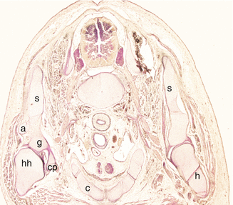

Fig. 2.3
Stage 19 (18 mm CRL) embryo transversally sectioned. Hematoxylin-Eosin stained. (×2). a acromion, c clavicle, cp coracoid process, g glenoid, h humerus, hh humeral head, s scapula
Each precartilaginous mass for the future clavicle undergoes independent ossification , the bone forming a central core to the precartilage; cartilage cells are seen in inner segment at this stage of development [6, 8, 9].
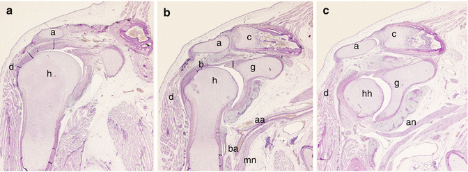
Stage 20 (18–22 mm; 50 days), stage 21 (22–24 mm; 52 days), stage 22 (23–28 mm; 54 days) and Stage 23 (27–31 mm; 56 days) (Figs. 2.4 and 2.5). From stage 20, the arterial pattern has already achieved its definitive morphology and during the 19 mm stage the two independent ossified clavicular centres fuse by a bony bridge resulting from the ossification of the precartilaginous mass. Between 24 and 27 mm the clavicular development finishes, both chondral and perichondrial bone are formed. Cartilage is now present in the hollow at the outer end of the outer segment, and the anlage of the deltoid tubercle is present [6].
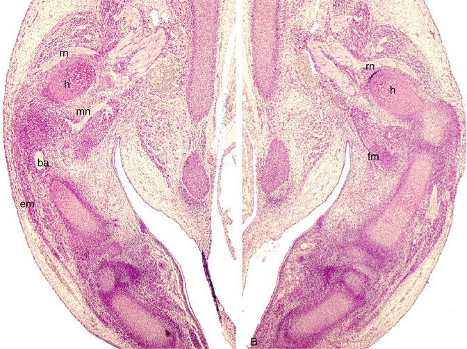
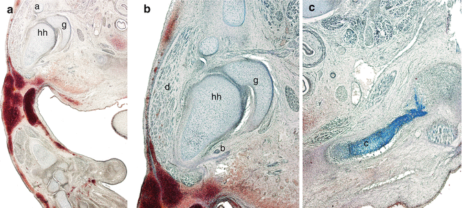

Fig. 2.4
Stage 20 (20 mm CRL) embryo transversally sectioned. Hematoxylin-Eosin stained. (×2). (a) Right upper limb. (b) Left upper limb. ba brachial artery, em extensor muscles, fm flexor muscles, mn median nerve, h humerus, rn radial nerve

Fig. 2.5
Stage 20 (27 mm CRL) embryo transversally sectioned. Azan stained. a (×2). b and c (×4). a acromion, b biccipital tendon, c clavicle, d deltoid muscle, g glenoid, hh humeral head
2.2.2 Fetal Development
Fetal CRL
30 mm (9 weeks), 53–58 mm (10–11 weeks) (Fig. 2.6). It has been described that the various components of the shoulder joint are discernable by 10 weeks. The head of humerus with its two tuberosities and intertubercular sulcus lodging the biceps tendon, acromion, coracoid process and spine of scapula are cartilagenous. Ossification occurs in the shaft which with due course of time extends higher up [9]. Periosteum of the humeral shaft has inner cellular and outer fibrous layers [2].
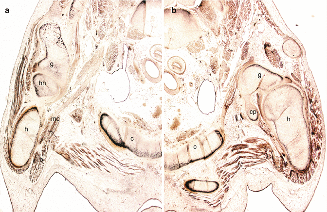

Fig. 2.6
Nine weeks development fetus (34 mm CRL) transversally sectioned. Bielchowsky stained. (a) Right upper limb (×2). (b) Left upper limb (×2). c clavicle, cp coracoid process, g glenoid, h humerus, hh humeral head, mc musculocutaneous nerve
The humeral head is very primitive and appears as a small protrusion. The glenoid labrum is a very thin bright rim attached to the glenoid margin. The tendon of the long head of biceps muscle looks small rounded band like a cord, passed over the humeral head, attached to the superior part of the glenoid labrum and separated from the humeral head by a small cavity. The joint cavity is still narrow and small and the capsule very thin. No ligaments can be detected at this age [10].
The scapula shows a concave glenoid fossa and the neck can be differentiated. The coracoid process is larger in size than the acromion, which is still cartilagenous. The joint cavity can be clearly visualized. The tissue lining the joint cavity is loose like synovial tissue and inferiorly it is reflected on the neck of humerus laterally and medially it is attached to glenoid labrum. The capsule is seen as continuation of perichondrium and is made of collagen fibres. It is more cellular than fibrous. Ossification of the humeral shaft progresses distal to the attachment of latissmus dorsi and teres major. The acromioclavicular joint can be recognized; it is lined by flattened cells. The acromial perichondrium extends to clavicle and serves the purpose of a capsular ligament. The lateral end of the clavicle is cartilagenous in nature [2].
The mesenchymal cells lining the articular surfaces and capsule of the shoulder joint are flattened and form a synovial membrane by 8–10 weeks while synovial villi develop by 11 weeks. The mesenchymal tissue surrounding the developing joint which is continuous with perichondrium forms a sleeve-like membrane which eventually transforms into a capsular ligament by 9 weeks. The capsule develops by 10 weeks and with increasing time the number of collagen fibres tends to increase. Coracohumeral and superior glenohumeral ligaments appear by the time of 10 weeks (Fig. 2.7).
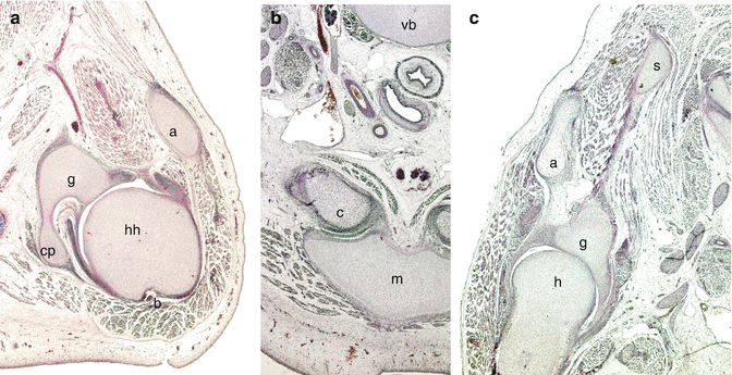

Fig. 2.7
Ten-eleven weeks development fetus (55 mm CRL) transversally sectioned. (a) Left shoulder (×2) Hematoxylin-Eosin stained. (b) Sternoclavicular joints level (×). Azan stained. (c) Right shoulder (×2). Azan stained. a acromion, b biccipital tendon, c clavicle, cp coracoid process, g glenoid, h humerus, hh humeral head, s scapule, m sternal manubrium, vb vertebral body
The rotator cuff covering the humeral head appears initially as an insertion of the infraspinatus at 9 weeks. Similar to the biceps long tendon, the tendons of the supraspinatus, infraspinatus, and subscapularis are located together outside the joint cavity and separated from it by a thick membranous structure, possibly the joint capsule, covered by a primitive glenohumeral ligament. This primitive glenohumeral ligament appears to be established as a transient, but complete collateral ligament. At 12 weeks, however, it becomes modified so that the rotator cuff tendons become attached to the humeral head [11] (Fig. 2.8).
