Fig. 26.1
Three parts of the deltoid . The anterior, middle, and posterior portions of the deltoid converge and run distally to the deltoid tuberosity on the humeral shaft
The origin and insertion of the deltoid have two attachment patterns: the tendinous attachment and the direct attachment of the muscle fibers. The muscle fibers of the anterior portion attach directly to the periosteum of the anterior surface of the distal clavicle. The muscle fibers of the posterior portion also attach directly to the periosteum of the scapular spine . In the middle portion, four intramuscular tendons originate from the lateral aspect of the acromion (Fig. 26.2a) [21, 28]. The muscle fibers arise from these intramuscular tendons and run downward to the intramuscular tendons of the insertion site. Tendinous insertion forms three discrete lines [14] or M-shaped insertion (Fig. 26.2b) [26, 28]. The anterior portion attaches to the anterior tendinous insertion and the posterior portion attaches to the posterior tendinous insertion. However, the middle portion diverges into three portions and each portion attaches to the anterior, middle, and posterior tendinous insertions, respectively. In other words, three origins and three insertions do not match with each other. The anterior insertion has three (A1, A2, and A3), the middle insertion has one (M1), and the posterior insertion has three (P1, P2, and P3) intramuscular tendons [19, 28].
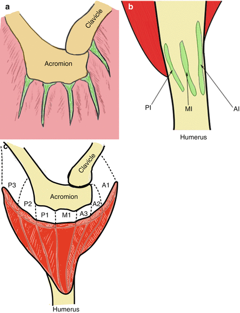
Fig. 26.2
Segments of the deltoid . The deltoid has several intramuscular tendons at its origin (a). The tendinous insertion of the deltoid forms three discrete lines (b). AI anterior insertion, MI middle insertion, PI posterior insertion (Modified from Rispoli et al. [26]). Based on the origins and insertions, the deltoid muscle can be divided into seven segments (c). The anterior segments (A1, A2, and A3) converge and attach to the anterior insertion. The middle segment (M1) attaches to the middle insertion. The posterior segments (P1, P2, and P3) converge and attach to the posterior insertion
According to the attachment pattern of the muscle fibers to the intramuscular tendons of the insertion, the deltoid can be divided into seven segments: i.e., A1, A2, A3, M1, P1, P2, and P3 segments (Fig. 26.2c) [28]. A1, A2, and A3 segments, M1 segments, and P1, P2, and P3 segments attach to the anterior, middle, and posterior insertions, respectively. In the classical division, A1 segment corresponds to the anterior portion, A2, A3, M1, and P1 segments correspond to the middle portion, and P2 and P3 segments correspond to the posterior portion. As the intramuscular tendons are clearly depicted on T2-weighted transverse magnetic resonance (MR) images with fat suppression, these seven segments can be differentiated on MR images [28, 37].
The tendon-muscle-tendon unit is known as the basic functional unit of the muscle, and thus, these anatomical segments based on the intramuscular tendons should be taken into consideration when the function of the deltoid is discussed.
26.2 Innervation
Innervation of the deltoid is supplied by the axillary nerve (C5 and C6) branched from the posterior cord of the brachial plexus. The axillary nerve passes through the quadrilateral space from anterior to posterior direction and splits into two branches (the anterior and the posterior branches) within the quadrilateral space. Anterior branch travels around the surgical neck of the humerus and supplies the middle and anterior portions of the deltoid (Fig. 26.3). Moreover, sub-branches from the anterior branch supplies the posterior portion [18, 35]. The posterior branch runs posteriorly and supplies the posterior portion of the deltoid. The posterior branch also sends a motor branch to the teres minor muscle and a sensory branch (superior lateral brachial cutaneous nerve) to the superolateral area of the shoulder called “regimental badge.” The posterior portion of the deltoid has double-supply from the anterior and posterior branches of the axillary nerve in 89.1 % of the cadaveric specimens [18]. In their series, the posterior portion is supplied by the anterior branch alone in 2.3 % and by the posterior branch alone in 8.5 % [18]. The mean diameter of the anterior and posterior branches are 4.0 and 3.3 mm, respectively [29]. The mean distance between the acromial edge and the axillary nerve varies among the reporters [20] reported that the distance from the humeral head and the axillary nerve ranged from 4.0 to 6.7 cm, however, the axillary nerve moves superiorly during abduction. The distance between the acromion and the axillary nerve ranged from 66.6 to 72.6 mm in the hanging arm position and from 53.9 to 61.6 mm in 60° of abduction [7].
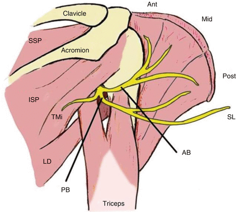
Fig. 26.3
Axillary nerve. The axillary nerve is divided into the anterior and the posterior branches in the quadrilateral space . The anterior branch innervates the whole deltoid , and the posterior branch provides a motor branch to the teres minor muscle and a sensory branch to the superolateral area of the shoulder. AA anterior branch of the axillary nerve , PA posterior branch of the axillary nerve , SL superior lateral brachial cutaneous nerve, ant anterior portion, mid middle portion, post posterior portion
Electromyographic assessment suggests that the deltoid consists of at least seven segments coordinated by the central nervous system [3, 4, 34]. However, these functional seven segments may differ from the anatomical seven segments divided by the intramuscular tendons. In other words, the relation between the innervated segments and the anatomical segments is still unclear and needs further investigation.
26.3 Vascularity
The vascular supply of the deltoid is largely derived from the posterior circumflex humeral artery, which travels with the axillary nerve through the quadrilateral space (Fig. 26.4). The posterior circumflex humeral artery supplies the middle and the posterior portions of the deltoid. The thoracoacromial artery also provides a supply to the deltoid muscle. The thoracoacromial artery is branched from the axillary artery and separates into two branches: the deltoid artery and the acromial artery. The deltoid artery runs near the deltopectoral groove and supplies the anterior portion of the deltoid [5]. The acromial artery travels in a deep layer of the deltoid near the clavicle and acromion and gives some branches to the middle portion. The anterior circumflex humeral artery sends a branch to the anterior portion in 63 % of cadaveric specimens [11].
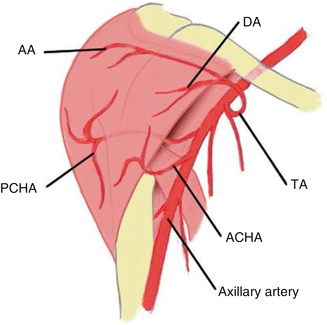
Fig. 26.4
The blood supply of the deltoid . The acromial and deltoid arteries, which branched from the thoracoacromial artery, and the anterior and posterior circumflex humeral arteries supply the deltoid muscle. TA thoracoacromial artery, AA acromial artery, DA deltoid artery, ACHA anterior circumflex humeral artery, PCHA posterior circumflex humeral artery
The venous branches accompany the arterial branches, except the cephalic vein , which runs in the deltopectoral groove.
26.4 Function
The motion of the shoulder is a complex of actions in many directions such as flexion/extension, abduction/adduction, internal rotation/external rotation. In addition, many muscles co-work in any single motion. Therefore, it is quite difficult to assess the participation of each muscle to the shoulder movement. The muscle function is evaluated by various methods, such as the physiological cross-sectional area (PCSA), the moment arm, the potential moment, and the electromyographic (EMG) activity [8, 15, 25, 33]. The deltoid has a largest PCSA in the shoulder girdle and is a key muscle in abduction of the shoulder. Moreover, the deltoid co-works with the rotator cuff muscles and constructs a force couple to move the shoulder joint smoothly.
The anterior portion of the deltoid elevates the arm forward with some contribution by the clavicular portion of the pectorals major and the biceps brachii. The middle portion elevates the arm laterally. The posterior portion works with the teres minor and the latissimus dorsi and elevates the arm backward [22]. In a moment arm study, the posterior portion was estimated to provide 14 % of the shoulder extension torque in addition to 20 % of the shoulder extension torque [8].
The deltoid used to be thought to push the humeral head superiorly during arm elevation. However, recent biomechanical studies have demonstrated that the deltoid also stabilizes the humeral head against the glenoid fossa during arm elevation [2, 23, 31] (Fig. 26.5). This function is very important in a rotator cuff deficient shoulder.
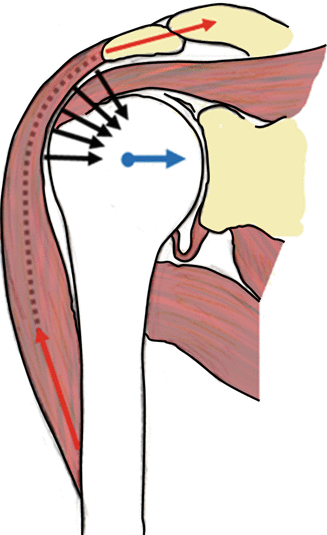
Fig. 26.5
The deltoid muscle as a stabilizer of the humeral head. The contraction force of the deltoid (red arrows) is converted into the stabilizing force (black arrows), which compresses the humeral head against the glenoid fossa (blue arrow). (Modified from Nyffeler et al. [23])
Biomechanical studies during the throwing motion have demonstrated that the deltoid acts as an anterior stabilizer along with the rotator cuff muscles at 90° of abduction and 90° of external rotation. Although this anterior stabilizing effect is stronger in 90° of elevation in the scapular plane than in the coronal plane, it is still weaker than the stability provided by the rotator cuff [13, 17].
26.5 Pathology of the Deltoid
Axillary nerve palsy is not uncommon, but the other conditions of the deltoid are rare.
26.5.1 Rupture of Deltoid Muscle
A spontaneous rupture of the deltoid muscle is uncommon and usually considered as a complication of the shoulder surgery. In case after acromioplasty, the deltoid origin is partially detached from the acromion . The open acromioplasty sacrifices 40 –70 % of the deltoid origin [30], whereas the arthroscopic acromioplasty preserves most of the footprint of the deltoid.
An isolated tear of the deltoid is reported in sports injuries and motor vehicle accidents. A tear occurs most frequently near the musculotendinous junction or at the origin from the acromion . A rupture at the middle of the muscle belly is rare [12]. A massive rotator cuff tear causes a deltoid tear, especially the middle portion. In a shoulder with a massive rotator cuff tear, the humeral head migrates superiorly and impinges against the acromial edge. At the same time, the undersurface of the deltoid muscle suffers a friction with the greater tuberosity, which may lead to a tear of the deep fascia of the deltoid. In a series of massive rotator cuff tears operated using a lateral para-acromial incision, the deltoid detachment occurred in 8 % of the patients during the first three months after surgery [9].
Related posts:

Stay updated, free articles. Join our Telegram channel

Full access? Get Clinical Tree


