Fig. 1.1
Appendicular skeleton of the earliest tetrapods (rhipidistian, bony fish), where the earlier connection to the skull was lost leading to the formation of the pectoral girdle and an increase of the mobility of the head. The cleithrum and clavicula are located more ventrally, the scapulocoracoid more dorsally; the interclavicle connects both the clavicles (Reprinted with permission from: Kardong [1])
1.2.2 Amphibians
In amphibians, with the acquirement of terrestrial habits, the tripartite type of pectoral girdle made its first appearance: the coracoid became segmented into the anterior procoracoid and posterior coracoid , while the clavicle formed a connection to the procoracoid [2].
1.2.3 Reptiles
In reptiles, the pectoral girdle has migrated caudally from its intimate relation to the head, as seen in fish. The reptilian pectoral girdle comprised a scapula, a clavicle, which replaced the provoracoid, and a coracoid.
The evolution of the procoracoid varies by species. It becomes part of the anterior scapula around the glenoid fossa; in others, it is fused with the sternum, or is replaced by the clavicle . Although the scapula and coracoid process are anatomically united, genetic patterning of the coracoid and the scapula is under the control of different Hox genes, lending further support to the view that each is a separate phylogenetic derivative [2].
1.2.4 Birds
The avian pectoral girdle became specialized to enable flight. The clavicles became essential in suspending the limb away from the body and the coracoid became large and strong in response to the muscular demands of flight. Consequently, the scapula was small, curved and narrow to allow greater motion. The keeled attachment for the strong pectoral muscles used in flight. The sternum became keel shaped to provide attachment for the strong pectoral muscles used in flight.
1.2.5 Mammals
The coracoid in mammals tends to become greatly reduced, forming an insignificant process on the scapula . The only other vestige of the bone is the coracoid ligament, extending from the coracoid process to the bone, in which may be found isolated masses of cartilage. This arrangement frees the scapula from any bone attachment to the skeleton. In mammals without clavicles, the scapula has no bony attachments whatsoever. It becomes the sole support for limb and provides attachments for muscles necessary for a freely movable extremity. New functional demands on the girdle resulted in the development of a projection of bone on the dorsal surface of the scapula , the spine, which extends downwards and ends in the acromion [2].
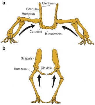
The clavicle undergoes changes during evolution, when in tetrapods a change in limb posture arises. In a sprawled posture, the forces are medially directed toward the shoulder girdle, conferring on medial elements a major role in resisting these forces: In these animals the clavicles are interconnected, a so-called interclavicle. As the limbs are brought under the body, these forces are directed less toward the midline and more in vertical direction, leading to a less prominent role of the clavicles (Fig. 1.2) [1].


Fig. 1.2
Change in the role of the shoulder girdle with change of limb posture. (a) Sprawled posture with medially directed forces to the shoulder girdle. (b) As limbs are brought under the body, these forces are more directed more ventrally. Especially, the role of the clavicle is diminished with these changes (Reprinted with permission from Kardong [1])
In mammals, which have acquired freedom of the forelimb to a marked degree, such as insectivores, primates and some marsupials and rodents, the clavicle is well developed. In others, including ungulates, carnivores and some rodents and marsupials, it is absent or rudimentary.
1.3 Phylogeny of the Shoulder Girdle: Musculature
Development of the shoulder and forelimb muscles in tetrapods comes from four sources: branchiomeric (jaw and pharyngeal muscles), axial, dorsal limb and ventral limb muscles (Fig. 1.3).


Fig. 1.3
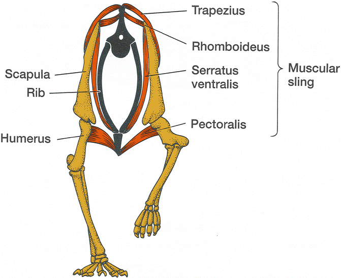
Muscular slingv of mammals. Some of the muscles arise from the branchiomeric muscles (trapezius ), some from the axial muscles (rhomboideus, serratus), and some from the ventral muscles (pectoralis ) (Reprinted with permission from Kardong [1])
1.3.1 Branchiomeric Muscles
Branchiomeric muscles: The branchiomeric muscles contribute the trapezius and mastoid (including the cleidomastoid and sternomastoid).
1.3.2 Axial Muscles
Axial muscles: The axial musculature contributes the levator scapulae, rhomboideus complex and serratus. These three muscles together with the trapezius form the muscular sling that suspends the body between the two scapular blades. As the shoulder girdle became separated from the skull, the branchiomeric and axial muscles developed into serving as part of the muscular sling through which the forelimbs are attached to the body.
1.3.3 Dorsal Muscles
Dorsal muscles: The dorsal muscles insert on the humerus and function to oscillate the humerus during movement or fix it in position, when an animal stands; of these muscles only the latissimus dorsi originates outside the limb from the body wall. The other dorsal muscles that act on the humerus are the teres minor , subscapularis and deltoideus, which may form two distinct muscles. The triceps is also derivative of the dorsal muscles but it acts to extend the forearm.
1.3.4 Ventral Muscles
Ventral muscles: The pectoralis is an important ventral muscle. In early mammals, the pectoralis consisted of four separate muscles, in primates only two remained: pectoralis major and pectoralis minor.
The ventrally positioned supracoracoideus in mammals originates dorsally on the lateral face of the scapula . The bony scapular spine divides this muscle into the supraspinatus and infraspinatus muscles, which insert on the humerus as well. The coracobrachialis from the coracoid runs along the front of the humerus.
In mammals, the biceps brachii has two heads, representing the fusion of two muscles, which insert into the forearm and are responsible for flexion.
1.3.5 Rototor Cuff
Rototor Cuff: Sonnabend et al. studied the rotator cuff muscles in 22 different animals, including marsupials, carnivores, ungulates and other primates. He identified rotator cuff muscles in all animals and that in the majority of animals the tendons inserted independently onto the tuberosities [3].
He observed that in some animals the tendons blended together to form a true rotator cuff (common tendon) in some primates (e.g. baboon and hominoids such as chimpanzee and orangutan). There was one marsupial species, the tree kangaroo, which formed a common tendon. All other animals in his study, the rotator cuff tendons were not interconnected.
There was a strong correlation of the presence of a true rotator cuff , with interconnected tendons, and the ability to perform activities overhead or away from the sagittal plane [3].
1.4 Comparative Anatomy: Tetrapods to Humans
Inman et al. have studied extensively the evolutionary changes of the shoulder girdle from tetrapodal primates through arboreal (tree living) primates to bipedal species, including hominoids [4].
1.4.1 Scapula
During evolution, the scapula changed in form when functional demands changed, including both change of posture prehensile tasks (form follows function!).
In reptiles, the scapula was broad and massive; however, with increased efficiency in locomotion, there were a number of changes. These included the following:
1.
Reduction in the size of the scapula
2.
Orientation of the glenoid changes from lateral to posterior-inferior
3.
Development of the scapular spine
4.
Disappearance of the procoracoid
5.
Reduction of size of coracoid process
6.
Rearrangement of muscles
The shape of the scapula is dependent on the posture: It is broad and massive in tetrapod animals that need a large serratus anterior muscle to support body weight. Alterations are brought about by change of posture from the pronograde (walking with the body horizontally) to the orthograde posture, as well as specialized requirements of a prehensile limb [2]. In orthograde posture, the position of the scapula changes from a lateral to a more dorsal position, also influenced by the flattening of the thorax. This change of position of the scapula also led to a lengthening of the clavicle .
Stay updated, free articles. Join our Telegram channel

Full access? Get Clinical Tree


