Vangsness (%)
Tuoheti (%)
Entirely posterior
22
28
Posterior dominant
33
55
Equal
37
17
Entirely anterior
8
0
Tuoheti et al. reported on 101 cadaveric shoulders macroscopically and then histologically found that the labral attachment of the long head of biceps tendon was posterior regardless of its macroscopic appearance, with 28 % entirely posterior, 55 % posterior dominant, and 17 % equal (Table 14.1). The variable macroscopic attachment pattern of the biceps tendon results from the differing attachment heights of the IGHL [57] Fig. 14.1.
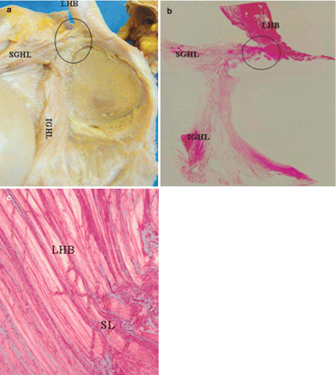
Fig. 14.1
(a) A posterior-dominant type. The LHB attaches to the SGT, mainly the posterior labrum. The IGHL attachment was high. The attachment of the MGHL was not clearly observed. (b) Histology (H & E) showed that the biceps attachment was entirely posterior. The IGHL linked to the SGHL on the superior labrum. (c) The polarized microscopic examination of the circled area in (b) showed that the fiber orientation of the biceps was totally posterior. SL superior labrum, LHB long head of biceps tendon, SGHL superior glenohumeral ligament, IGHL inferior glenohumeral ligament (Used with permission [57])
The LHB slides passively on the humeral head during abduction and rotation. It slides up to 18 mm in and out of the glenohumeral joint during forward flexion and internal rotation Fig. 14.2.

Fig. 14.2
LHB and humeral abduction. (a) Humeral abduction, the biceps tendon is stabilized by the transverse ligament. (b) The transverse ligament can impinge on the biceps tendon, producing an hour-glass constriction of the tendon. (c) Humeral adduction, LHB slides on the mobile humeral head up to 18 mm from forward flexion and internal rotation compared to neutral (Copyright Dr Gregory Bain), (d) The clinical photo demonstrates a spur and tendon abrasion (Copyright Di Giacomo [22])
Because of its location, the LHB is exposed to extraarticular constraints from possible subacromial impingement, and intraarticular restriction from the constant sliding of the tendon within the bicipital groove during elevation and rotation of the shoulder [1].
The LHB has an intraarticular and an extraarticular portion. The intraarticular portion is extrasynovial and is essentially static within the joint as the groove slides over the biceps during abduction and rotation [31]. This portion is flat and wide in shape with a length of 34.4 ± 4.2 mm.
The extraarticular portion is round and narrower with a length of 30.6 ± 5.7 mm [59]. The bicipital groove is an hourglass-shaped corridor between the greater and lesser tuberosities; this groove is narrowest and deepest at its midportion [47].
The reported approximate cross-sectional area of the tendon is
The contribution of the superior glenohumeral ligament (SGHL) and coracohumeral ligament (CHL) to the “biceps pulley” mechanism is critical to the stability of the tendon, with the failure of this mechanism leading to instability [1].
14.2.1 Biceps Restraints
The contours of tuberosities and a pulley mechanism formed by soft tissues maintain the biceps in its groove. There are two pulleys:
Medial (inferior or reflection pulley), formed by CHL, SGHL, and the superior border of the subscapularis
Lateral (superior pulley), formed by the anterior border of the supraspinatus and secondarily the rotator cuff cable Fig. 14.3
Fig. 14.3
Medial (inferior or reflection pulley) and lateral (superior pulley)
There exists a close relation among the LHB, the superiormost intramuscular tendon of the subscapularis muscle, and the SGHL [4] See Fig. 14.4.
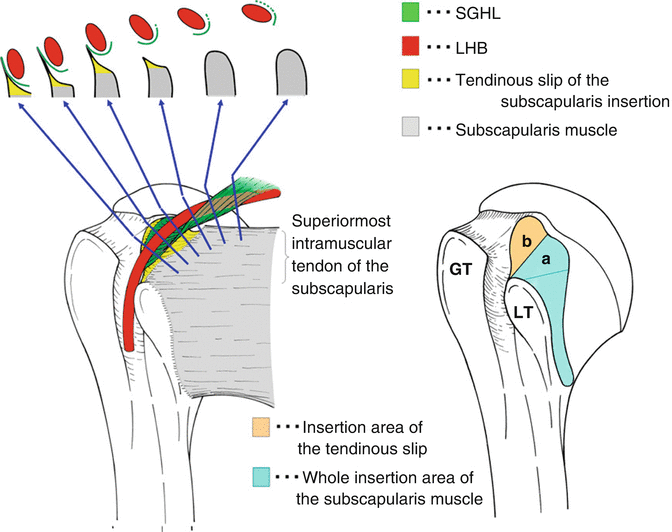
Fig. 14.4
Close relations of the long head of biceps tendon. Diagram of the relationships of the LHB (red), subscapularis (gray), and the SGHL (green). The intramuscular tendon of the subscapularis inserts into the superior footprint on the lesser tuberosity (area a). This insertion sends a thin tendinous slip to the fovea capitis of the humerus (area b). The SGHL passes from its glenoid attachment, passes laterally and wraps around the LHB in a spiral fashion, and attaches to the tendinous slip of the subscapularis insertion. The superior subscapularis tendon and the SGHL stabilize the LHB. The CHL and SGHL are continuous, and therefore also provide some stability for the LHB. GT Greater tuberosity, LT lesser tuberosity, LHB long head of the biceps tendon, SGHL superior glenohumeral ligament, CHL coracohumeral ligament (Used with permission [4])
14.2.2 Irrigation
The LHB blood supply is from the brachial artery. Three arteries supply blood to the bicipital tendon [17]. Blood supply to the proximal part of LHB is from the anterior circumflex artery, with the branches running along the bicipital groove in both cranial and caudal directions. There is a characteristic vascular pattern on the superficial surface of the tendon within the groove, while the deep “sliding” surface is avascular and composed of fibrocartilage [1]. Labral branches from the suprascapular artery supply the proximal tendon [21].
The distal portion of the tendon receives branches from the deep brachial artery [17] Fig. 14.5.
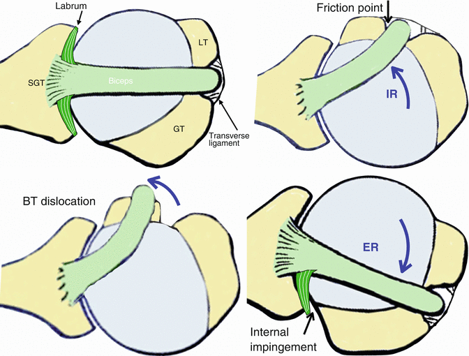
Fig. 14.5
LHBT and humeral rotation. (a) Medially, the LHBT is stabilized to the (SGT) supraglenoid tubercle, with extensions to the posterior +/− anterior labrum. Laterally, it is stabilized by the transverse ligament. Anterior and posterior stabilities are provided by the anteromedial (AM) and posterolateral (PL) pulleys. Each pulley has an osseous, ligamentous (static), and dynamic component. AM pulley- LT, subscapularis, SGHL. PL pulley- GT, supraspinatus. (b) Internal rotation of the humeral head will create a friction point, and the LHBT will abrade over the superior lesser tuberosity and superior subscapularis tendon. Note the AM pulley is dynamically supported by the subscapularis tendon. (c) Failure of the anterior pulley will produce a biceps tendon instability +/− subscapularis tendon tear. (d) With external rotation, especially with abduction (“cocking position”), there will be internal impingement, due to the posterior superior labrum abutting against the greater tuberosity and adjacent rotator cuff (Copyright Dr Gregory Bain)
14.2.3 Innervation
Clinically, the LHB has been considered a pain generator in the shoulder. According to Alpantaki et al. [2], a rich innervation through a network of sensory sympathetic fibers may explain the bicipital pain. This pattern of innervation is asymmetrical, more concentrated at the biceps origin and less so at the musculotendinous junction. The musculocutaneous nerve is responsible for the motor innervation of the muscle.
14.2.4 Anatomical Variations
Dierickx et al., in a review of 3,000 cases of shoulder arthroscopies, found 57 cases (1.91 %) with variations: the simple vinculum or pulley-like sling, the partial or complete mesotenon between the biceps and the capsule, the completely adherent LHB, the double tendon origin, the reversed-type split tendon, and the complete absence of the LHB. They suggested a classification of 12 variations of the intraarticular portion of the LHB [23].
Four major families, each divided into 1–5 subgroups, were described: Fig. 14.6
1.
Synovial mesentery (vinculum, pulley-like sling)
2.
Adherent to rotator cuff
3.
Split or bifid tendon
4.
Absence of LHB
Many of these variations, such as the vinculum, the pulley-like sling, and the mesotenon-like adhesions, are mostly of a common kind. They refer to the human embryological evolution of the LHB tendon, which migrates intraarticularly after the fourth month of gestation. This process may apparently stop prematurely, which results in one of the mentioned conditions. These adhesions probably do not cause any pathology [23].
14.2.5 Comparative Anatomy
Comparative anatomy has demonstrated the evolutionary movement of the scapula to a more frontal plane with associated torsion of the humeral shaft, thus reducing the action of the LHB at the shoulder [15, 31, 35, 39].
Decreased retroversion of the proximal humerus has led to the groove no longer being centered in the plane of the humeral head, but lying at an angle of approximately 30°. As a consequence, the LHB is forced to bear on the lesser tuberosity at the medial wall of the groove, instead of at its middle. Such a position renders the tendon vulnerable to pathology, since the lesser tuberosity itself acts as a pulley [1].
14.3 Function
The function of the biceps tendon has been one of debate. Some suggest that it has no role and is only a vestigial structure, being the “appendix of the shoulder.” Its role in glenohumeral kinematics is controversial. It is important for elbow function, as it is the prime forearm supinator and a significant elbow flexor.
For the shoulder, cadaveric studies have reported that it has a stabilizing effect on the glenohumeral joint in all directions. It is a humeral head depressor and is also an abductor in external rotation.
Despite the possible kinematic effect of the biceps tendon, in vivo studies are yet to establish its stabilizing effect on the shoulder. We know there is minimal functional impact of a biceps tenotomy or tenodesis. Furthermore, EMG studies have demonstrated little or no activation of LHB when the elbow is immobilized. During throwing, the biceps has been shown to function predominantly as an elbow flexor.
In summary, while important for elbow function, the biceps tendon has only a minor contribution to shoulder function. However, if a biceps lesion exists, it is often painful, and often has a significantly detrimental effect on shoulder function.
14.4 Imaging Study
Ultrasound has proven to be an accurate diagnostic method for LHB tears; however, it is less effective when studying other disorders, such as inflammatory changes or partial tears [52].
Magnetic resonance imaging (MRI) is useful for proximal biceps pathology, both for articular and extraarticular portions. Increased fluid in the synovial sheath is suggestive of tenosynovitis. In sagittal and coronal views, a hyperintense signal under the superior labrum is suggestive of injury of the labrobicipital insertion. The presence of paralabral cysts adjacent to the labrobicipital insertion is also suggestive of injury. The use of gadolinium-enhanced MRI increases diagnostic accuracy; however, experienced radiologists must analyze it so as not to confuse normal anatomic variants such as the sublabral recess with pathological injuries [43]. The diagnostic accuracy of labrobicipital injuries is increased by abduction and external rotation of the arm, as it simulates the peel-back phenomenon, showing the medial labrobicipital complex caudal to the glenoid articular plane Fig. 14.7 [13]. The normal and pathologic anatomy of the biceps reflection pulleys may also be studied by MR arthrography. Oblique sagittal images and axial images are valuable for identifying the individual components of the pulley system [46].
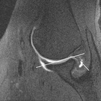
Fig. 14.7
MR arthrogram image in ABER, showing displacement of the posterosuperior labrum (arrow) medial/caudal to the glenoid articular plane (line) (Reproduced from Borrero et al. [13])
14.5 Arthroscopic Biceps Anatomy
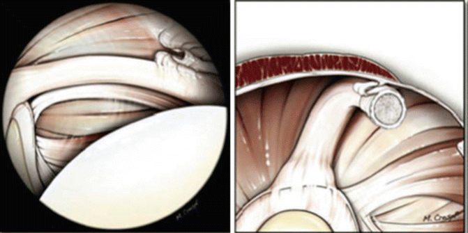
The LHB tendon is one of the most important reference points for orientation during the glenohumeral arthroscopy. The arthroscopic view starts from the posterior viewing portal, where its intraarticular portion and its origin at the supraglenoid tubercle by the labrobicipital union are easily visible. With the arm at the side and in neutral rotation, a better visualization of the intraarticular portion is obtained. It is of major importance to assess the LHB extraarticular or intertubercular portion by applying downward traction with a probe, with an additional 3–5 cm view able to be obtained [26]. The extraarticular biceps portion is a common location for “lipstick synovitis,” delamination, and partial tears. The medial and lateral pulleys complex can be seen with the scope from the posterior portal with the arm in 30° flexion and neutral rotation Figs. 14.8 and 14.9. Associated tears of the subscapularis tendon are common (Fig. 14.8b). Medial displacement of the LHB with internal rotation suggests an anteromedial pulley injury, whereas lateral displacement with external rotation of the arm suggests a posterolateral pulley injury. This is the so-called swinging test [9, 14].
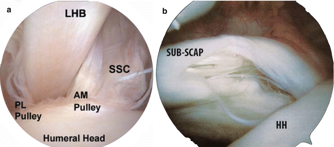
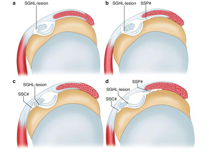
Fig. 14.8
(a) Left shoulder. Arthroscopic view of anteromedial (AM) pulley and the posterolateral pulley from posterior viewing portal. (b) Partial tear of the superior border of the right subscapularis tendon
Fig. 14.9


Fig 14.9: Habermeyer LHB instability due to pulley lesions. (a) Group 1: isolated lesion of the SGHL (arrow). (b) Group 2: SGHL lesion and partial articular-side supraspinatus tendon tear (SSP#) (arrows). (c) Group 3: SGHL lesion and tear of the upper third of the subescapularis tendon (SSC#) (arrows). (d) Group 4: combined lesion of the SGHL, a partial articular tear of the supraspinatus (SSP#) and a tear of the upper third of the subescapularis tendon (SSC#) (arrows). (Reproduced from Habermeyer et al. [30])
Related posts:


Stay updated, free articles. Join our Telegram channel
Full access? Get Clinical Tree
