Fig. 17.1
Left clavicle (inferior view). (a) Attachments of the conoid and trapezoid ligaments. (b) Relative position of the coronoid process. (c) Cadaveric attachment sites
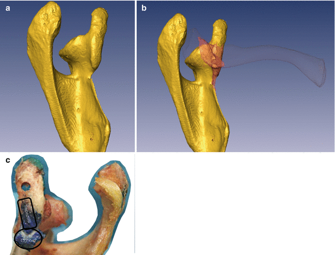
Fig. 17.2
Left scapula with coracoid process (superior view). (a) Attachments of the conoid and trapezoid ligaments. (b) Relative position of the clavicle. (c) Cadaveric attachment sites
Although 1 % of people have coracoclavicular bar or joint, AC joint is the only articulation between clavicle and scapula [8]. In spite of many anatomical variations, AC joint is a synovial type of planar diarthrodial joint (Fig. 17.3). Although variable the hyaline cartilage covered convex and oval facet on anterior portion of the distal clavicle articulate with the concave small facet-like portion of the anteromedial aspect of the acromion process. The mean size of the joint is 9 × 19 mm in adults. The joint line is oblique and slightly curved permitting the protraction and retraction of the scapula [8] (Figs. 17.4, 17.5, 17.6, and 17.7).
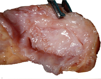
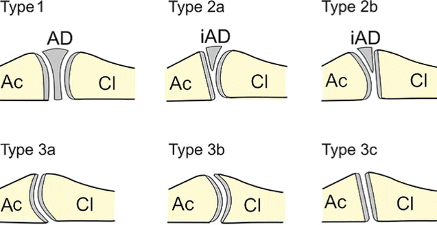
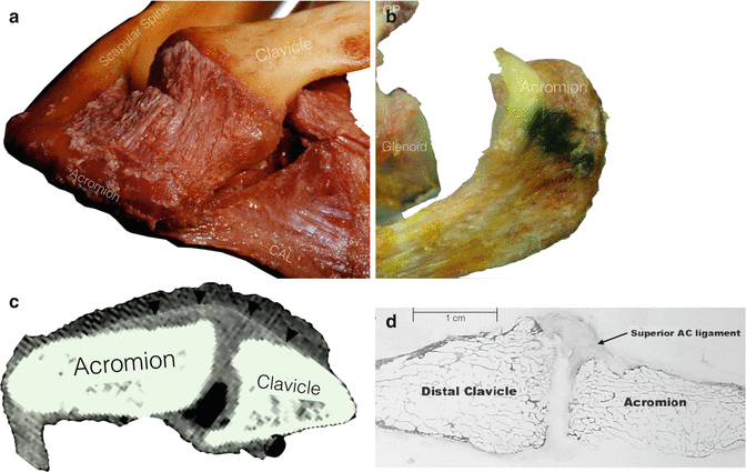
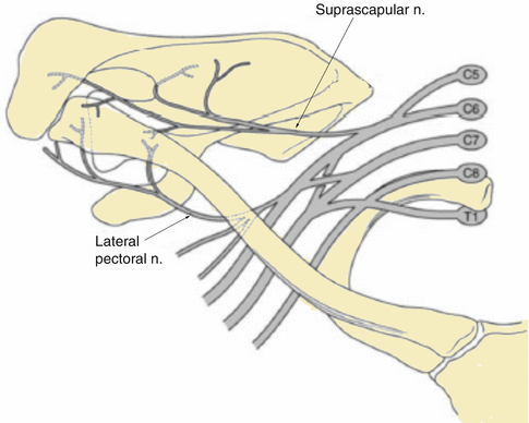
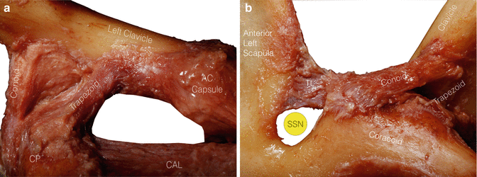

Fig. 17.3
Lateral clavicle articulation (lateral view). Lateral articulation of the clavicle demonstrating that the AC joint is a synovial planar diarthrodial joint, which is covered with variable hyaline cartilage

Fig. 17.4
Types of AC joints. Type 1: double ellipsoid joint (4 %). The articular disk (wedge shape) completely divides the articular cavity. It is attached to the articular capsule at its periphery. Both articular surfaces are slightly convex. Type 2: incomplete articular disk incompletely divides the AC joint cavity (25 %). Type 2a: clavicle facet is convex and the acromion flat. Type 2b: clavicle facet is flat and the acromion convex. Although the articular surfaces were incongruent, the incomplete disk compensates. Type 3: absent articular disk (71 %). Type 3a: clavicle facet convex and acromion facet concave- an ellipsoid joint. Type 3b: clavicle facet concave and acromion facet convex. Type 3c: both articular surfaces flat – planar joints

Fig. 17.5
Superior AC ligament. The superior AC capsular ligament attaches close the AC joint articulation. (a) Cadaveric view of superior capsule. (b) Cadaveric site of insertion of the superior AC joint ligament. (c) CT scan. (d) Histology

Fig. 17.6
Nerve supply of the AC joint. The AC joint is supplied by the Suprascapular, axillary and lateral pectoral nerves

Fig. 17.7
Gross anatomy of coracoclavicular ligaments. (a) Anterior view. (b) Anterior medial view. CP coracoid process, TL transverse ligament, SSN suprascapular nerve, CAL coracoacromial ligament
A fibrocartilaginous disk cushions the joint, corrects for incongruencies, and acts in a load-bearing fashion similar to the meniscus in the knee [9] but others have attributed negligible function to it. This is composed of 75 % water, 20 % collagen (90 % type I), and 5 % proteoglycans, elastin, and other cells [10]. Variable inclinations exist, with being nearly vertical to angled downward and medially accounting for up to 50° [11]. Degeneration of the intra-articular disk, commonly observed in patients over the age of 50 years, begins as early as the timesfibers were confluent with the inferior AC ligament (ACL) at the acromial insertion. The small distance from the medial acromial articular surface to the beginning of the coracoacromial ligament (mean, 3.5 mm) stresses the close proximity of the coracoacromial ligament to the capsular insertions on the anteroinferior acromial surface, which can inadvertently be taken down during distal clavicle resection or co-planing. On the superior side of the AC joint, the trapezius was found to be confluent with the posterosuperior AC ligaments [13].
Barber et al. found no long-term instability after co-planing or hemi-resection (58 patients) of the AC joint or distal clavicle excision (23 patients) in a study in which all patients required resection of the inferior AC capsule and ligament. This illustrates the importance of maintaining the superior and posterior structures to ensure stability [15].
3-D CT scan showed [16] significant variability of the bone shape and size near the AC joint. ACJ subtends mean angle of 51° in the axial plane and 12° in the coronal plane with respect to the clavicular shaft. Hence distal clavicle resections should respect these angles and address the unique morphology of each AC joint to provide a symmetric bone resection without disruption of the AC joint capsule ACL. They proposed this anatomic-based recommendation of 5–7 mm of total resection (combining acromial and distal clavicle resection) should be adequate to provide relief of symptoms and likely to provide a more reliable outcome than larger resections.
Surgically important stabilizer of AC joint is extrinsic coracoclavicular ligaments (CCL) (conoid and trapezoid ligaments). Rather than resisting the traumatic displacements, these ligaments function to control and guide the AC joint movements and provide stability by complex interplay with AC joint capsule, AC ligament and dynamic stabilizers like deltoid, trapezius, and coracoids muscles. The CCL were determined to be greater than 3 times stiffer than the AC ligament. The AC joint compression loads can only occur when the distal clavicle is intact.
CCLs are responsible for suspending the scapula and the upper extremity from the under surface of the clavicle. They are the stronger, more vertically oriented ligaments. They arise from superior surface of coracoid posterior to the pectoralis minor attachment. The mean length of CCL is 19.4 mm. Conoid and trapezoid components are functionally and anatomically distinct and separated by a bursa between them. The conoid ligament thick and triangular, posteromedial in location, with short stout fibers almost vertical and insertion ends approximately 30 mm (females 28.9, males 33.5) from the joint line (Fig. 17.8). The trapezoid ligament is broad, thin, quadrilateral shape, and anterolateral in location. The insertion ends at mid-arc of the lateral curve and trapezoid ridge approximately 16 mm (females 16.1, males 16.7) from the ACJ line [17]. Average vertical height of coracoclavicular space is 1.1–1.3 cm [14]. The CCL strengthens the ACJ and mediate the synchronous scapuloclavicular rotation and scapulohumeral movement.
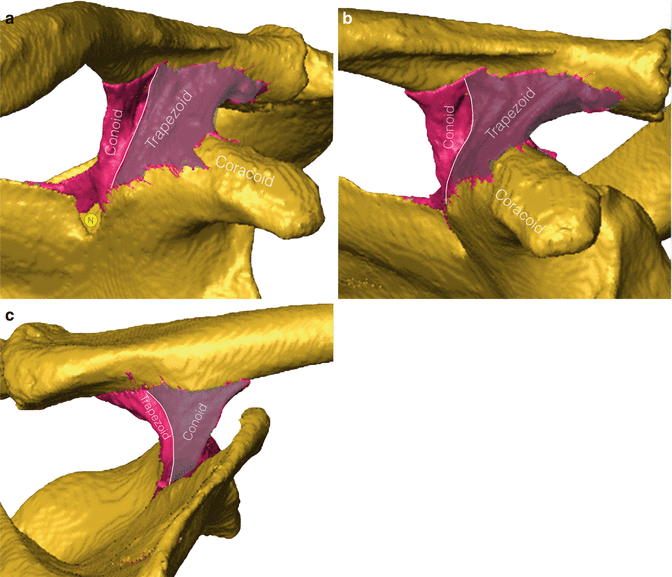

Fig. 17.8
Costoclavicular ligaments. Images demonstrate the relative position of the trapezoid and conoid ligaments: (a) Anterior view. (b) Anteriolateral view. (c) Aosteromedial view
The manner of attachment provides a mechanism for producing increased external rotation of the scapula. With the elevation of the humerus, scapula rotates to displace the coracoids inferiorly. The resulting tension in the CCL acts on the lateral curve to rotate the clavicle on its long axis. The crank-like phenomenon provided by the coracoclavicular ligaments and the S shape of the clavicle will not restrict the abduction of the arm.
The majority of anteroposterior stability (90 % resistance to anterior translation of scapula on clavicle) and distraction (91 % resistance to distraction) of the ACJ is provided by ACL. Most of the vertical stability (77 % of resistance to inferior translation of the scapula) is conferred by CCL. The conoid ligament is the primary restraint against anterior and superior loading, whereas the trapezoid ligament is the primary restraint against posterior loading. About 75 % of resistance to compression of ACJ is provided by trapezoid ligament [18].
The difference in the contributions by the two ligaments is most likely due to their relative orientations. In situ forces in each ligament are affected by coupled motions that occur during loading. This soft tissue force is redistributed during loading when a greater number of degrees of freedom of motion are allowed [19, 20].
The literature has supported the concept that the AC joint capsule is integral in maintaining normal joint contact and primarily resists motion in the AP (horizontal) plane, and the CCL primarily resist motion in the superoinferior (vertical) direction [21].
Fukuda et al. [18] quantified “the displacement as a function of the ligamentous constraints.” From selective ligament section studies they reported that with small displacements the acromioclavicular ligaments are the primary restraints to posterior (89 %) and superior (68 %) translation of the clavicle. With larger displacement, the conoid ligament was found to be the primary restraint (62 %) to superior translation. The trapezoid ligament was found to be the primary restraint to compression of the acromioclavicular joint at both small and large displacements. Hence the overriding of AC joint is resisted by the trapezoid ligament . Hence small displacement is limited by ACL, but large displacements are resisted by the CCL. Lee and coworkers further determined that the trapezoid ligament was the primary restraint to posterior displacement of the distal clavicle with an intact AC joint [22].
Stay updated, free articles. Join our Telegram channel

Full access? Get Clinical Tree


