The surgical anatomies of the two bones of the forearm differ significantly. The ulna has a subcutaneous border that extends for its entire length; the bone can be reached simply and directly without endangering other structures. In contrast, the upper two thirds of the radius are enclosed by a sheath of muscles. All surgery in the upper third of the radius is complicated further by the posterior interosseous nerve, which winds spirally around the bone close to, if not in contact with, its periosteum.
Three surgical approaches to the forearm are described in this chapter, all of which allow for the complete exposure of bone. In nearly every case, only part of the approach is required. The anterior approach to the radius is one of the classic extensile approaches, relying on subperiosteal dissection for protection of the posterior interosseous nerve. The posterior approach to the radius also makes use of an internervous plane, but still requires identification and preservation of the posterior interosseous nerve. The approach to the ulna cuts directly onto its subcutaneous border. The anatomy of the anterior approach to the radius, the approach to the ulna, and the anatomy of the posterior compartment of the forearm are considered separately. Because of the critical importance of the posterior interosseous nerve, its course is described in both anatomic sections.
Anterior Approach to the Radius
The anterior approach offers an excellent, safe exposure of the radius, exposing the entire length of the bone. Exposing the proximal third of the radius endangers the posterior interosseous nerve. By stripping the supinator muscle off the radius subperiosteally and using it to protect the nerve, however, the anterior approach avoids this danger. Still, great care must be taken in positioning retractors, because the nerve actually may touch the bone at the level of the distal portion of the neck of the radius, opposite the bicipital tuberosity, and posteriorly placed retractors can compress it against the bone. The approach first was described by Henry, and his name usually is associated with it.1
The uses of the anterior approach include the following:
Open reduction and internal fixation of fractures2
Bone grafting and fixation of fracture nonunions
Radial osteotomy
Biopsy and treatment of bone tumors
Excision of sequestra in chronic osteomyelitis
Anterior exposure of the bicipital tuberosity
Treatment of compartment syndrome
This section describes an approach that exposes the entire length of the bone. Ordinarily, only a portion of the approach is required.
Position of the Patient
Place the patient supine on the operating table, with the arm on an arm board. Place a tourniquet on the arm, but do not exsanguinate it fully before inflating the tourniquet. Venous blood left in the arm makes the vascular structures easier to identify. Finally, supinate the forearm (Fig. 4-1).
Landmarks and Incision
Landmarks
Palpate the biceps tendon, which is a long, taut structure that crosses the front of the elbow joint just medial to the brachioradialis muscle.
Palpate the brachioradialis, which is a fleshy muscle that arises with the extensor carpi radialis longus and brevis muscles from the lateral epicondyle of the elbow. The three muscles form a “mobile wad” of muscle that runs down the lateral aspect of the supinated forearm.
Palpate the styloid process of the radius. Note that this bony process is truly lateral when the hand is in the anatomic (supinated) position. The styloid process is the most distal part of the lateral side of the radius.
Incision
Make a straight incision from the anterior flexor crease of the elbow just lateral to the biceps tendon down to the styloid process of the radius. The length of the incision depends on the amount of bone that needs to be exposed (Fig. 4-2).
P.149
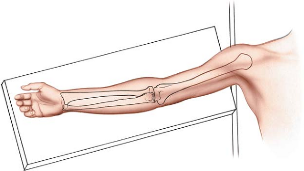 |
Figure 4-1 Position of the patient on the operating table, for the anterior approach to the radius. |
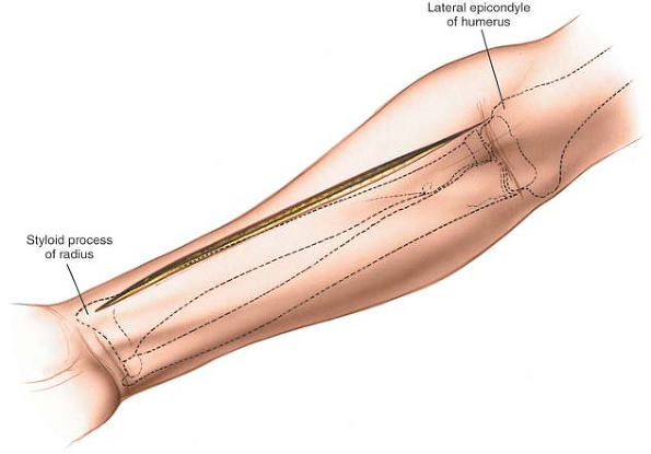 |
Figure 4-2 Make a straight incision on the anterior part of the forearm, from the flexor crease on the lateral side of the biceps down to the styloid process of the radius. |
P.150
 |
Figure 4-3 Internervous plane. The plane lies between the brachioradialis (radial nerve) and the flexor carpi radialis (median nerve). |
Internervous Plane
Distally, the internervous plane lies between the brachioradialis muscle, which is innervated by the radial nerve, just proximal to the elbow joint, and the flexor carpi radialis muscle, which is innervated by the median nerve (Fig. 4-3). Proximally, the internervous plane lies between the brachioradialis muscle, which is innervated by the radial nerve, and the pronator teres muscle, which is innervated by the median nerve.
Superficial Surgical Dissection
Incise the deep fascia of the forearm in line with the skin incision. Identify the medial border of the brachioradialis as it runs down the forearm, and develop a plane between it and the flexor carpi radialis distally. More proximally, the plane lies between the pronator teres and brachioradialis muscles (Fig. 4-4). Note that the medial border of the brachioradialis is surprisingly far across the forearm. At the level of the elbow the brachioradialis extends almost halfway across the forearm.
Begin dissection distally and work proximally. Identify the superficial radial nerve running on the undersurface of the brachioradialis and moving with it. The brachioradialis receives a number of arterial branches from the radial artery (called the recurrent radial artery) just below the elbow joint. Ligate this recurrent leash of vessels (Fig. 4-5). Take care to ligate these vessels and not avulse them, as avulsion is a potent cause of postoperative hematoma formation. Many vessels are present and all will need to be ligated and divided to allow the brachioradialis to be mobilized laterally.
The radial artery lies beneath the brachioradialis in the middle part of the forearm; therefore, it is quite close to the medial edge of the wound. It runs with its two venae comitantes, which remain prominent if the limb is not exsanguinated before the tourniquet is applied. Often, the artery may have to be mobilized and retracted medially to achieve adequate exposure of the deeper muscular layer, particularly at the upper and lower ends of the approach (see Fig. 4-5).
The superficial radial nerve, which is a sensory nerve in the forearm, also runs under cover of the brachioradialis muscle. Preserve the nerve, because damage to it may create a painful neuroma at the operative site (see Fig. 4-5). It is retracted laterally with the brachioradialis muscle.
P.151
 |
Figure 4-4 Incise the fascia and develop the plane between the brachioradialis and the flexor carpi radialis. |
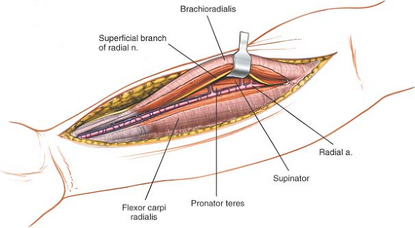 |
Figure 4-5 A leash of vessels from the radial artery supplies the brachioradialis. The vessels must be ligated to mobilize the brachioradialis laterally. Retract the superficial branch of the radial nerve with the brachioradialis muscle. |
P.152
Deep Surgical Dissection
Proximal Third
Follow the biceps tendon to its insertion into the bicipital tuberosity of the radius. Just lateral to the tendon is a small bursa; incise the bursa to gain access to the proximal part of the shaft of the radius. Because the radial artery lies superficial and just medial to the tendon at this point, deepen the wound on the lateral side of the biceps tendon (Fig. 4-6).
The proximal third of the radius is covered by the supinator muscle, through which the posterior interosseous nerve passes on its way to the posterior compartment of the forearm.
The posterior interosseous nerve is the single most important structure left vulnerable by this approach. To displace the nerve laterally and posteriorly (away from the surgical area), fully supinate the forearm, exposing, at the same time, the insertion of the supinator muscle into the anterior aspect of the radius (Fig. 4-7).
Next, incise the supinator muscle along the line of its broad insertion. Ensure that the muscle is detached by dividing its insertion and not by splitting the muscle. Continue subperiosteal dissection laterally, stripping the muscle off the bone (see Fig. 4-7). Lateral retraction of the muscle lifts the posterior interosseous nerve clear of the operative field, but be careful! Excessive traction may cause a neurapraxia of the nerve, and it recovers very slowly, taking up to 6 to 9 months. Finally, do not place retractors on the posterior surface of the radial neck, because they may compress the posterior interosseous nerve against the bone in patients whose nerve comes into direct contact with the posterior aspect of the radial neck (about 25% of all patients).3
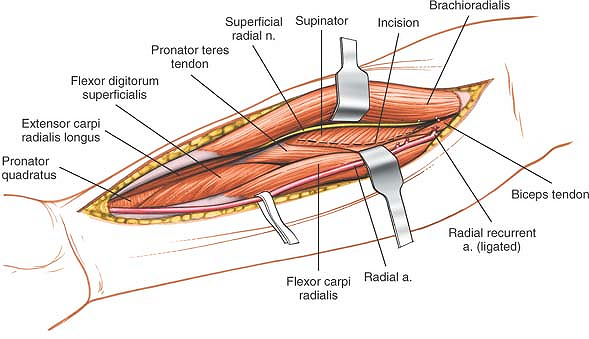 |
Figure 4-6 Deep to the brachioradialis and the flexor carpi radialis are the supinator muscle, the pronator teres, the flexor digitorum superficialis, and, most distally, the pronator quadratus. |
Middle Third
The anterior aspect of the middle third of the radius is covered by the pronator teres and flexor digitorum superficialis muscles. To reach the anterior surface of the bone, pronate the arm so that the insertion of the pronator teres onto the lateral aspect of the radius is exposed (Fig. 4-8; see Fig. 4-6). Detach this insertion from the bone and strip the muscle off medially. Preserve as much soft tissue as you can compatible with accurate reduction and fixation of the fracture. This maneuver detaches the origin of the flexor digitorum superficialis from the anterior aspect of the radius as well (Fig. 4-9).
Distal Third
Two muscles, the flexor pollicis longus and the pronator quadratus, arise from the anterior aspect of the distal third of the radius. To reach bone, partially supinate the forearm and incise the periosteum of the lateral aspect of the radius lateral to the pronator quadratus and the flexor pollicis longus. Then, continue the dissection distally, retracting the two muscles medially and lifting them off the radius (Fig. 4-10).
P.153
 |
Figure 4-7 With the patient’s arm in the supinated position, resect the origin of the supinator. Reflect the muscle laterally. Leave the posterior interosseous nerve in the muscle’s substance. The radial nerve enters the supinator through the arcade of Frohse (inset). Turning the forearm upward moves the nerve laterally, away from the operative field. The origin of the supinator muscle is easier to identify if the surgeon stays lateral to the biceps tendon and locates the bursa between it and the supinator. |
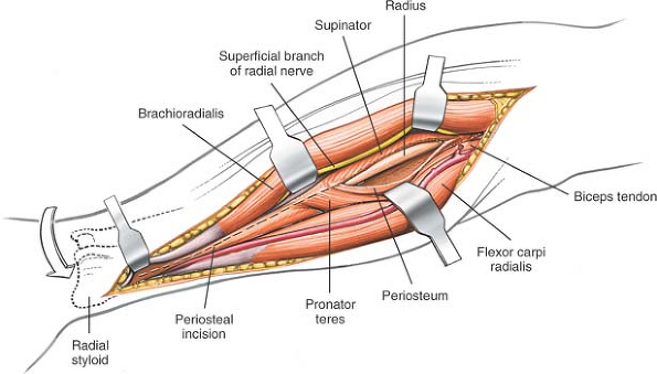 |
Figure 4-8 Turn the arm downward to identify the pronator teres muscle. Resect it along its insertion on the lateral aspect of the radius. |
P.154
 |
Figure 4-9 Continue dissection distally to uncover the distal part of the radius. Leave the periosteum intact. |
 |
Figure 4-10 With the arm in partial supination, remove the flexor pollicis longus and the pronator quadratus from the bone to expose the entire radius from its proximal to distal end. |
P.155
Dangers
Nerves
The posterior interosseous nerve is vulnerable as it winds around the neck of the radius within the substance of the supinator muscle. The key to ensuring its safety is to detach correctly the insertion of the supinator muscle from the radius. The insertion of the muscle is exposed completely only when the arm is supinated fully. Once the subperiosteal dissection is begun, the nerve is comparatively safe, but overzealous retraction still can lead to a neurapraxia (see Figs. 4-7, inset, and 4-13).
The superficial radial nerve runs down the forearm under the brachioradialis muscle. It becomes vulnerable when the “mobile wad” of three muscles is mobilized and retracted laterally (see Fig. 4-5). The superficial radial nerve is vulnerable to neurapraxia if it is retracted vigorously. Take great care, therefore, when retracting the nerve and warn your patients preoperatively that temporary paresthesia in the distribution of the superficial branch of the radial nerve may occur in the early postoperative phase.
Vessels
The radial artery runs down the middle of the forearm under the brachioradialis muscle. It is vulnerable twice during the anterior approach to the radius:
During mobilization of the brachioradialis. Protection depends on recognizing the artery. Its two accompanying venae comitantes are the best surgical guide, because the artery is surprisingly small after a tourniquet has been used (see Fig. 4-5).
In the proximal end of the wound, as the artery passes to the medial side of the biceps tendon. Damage to the artery at that level can be avoided by remaining lateral to the tendon (see Fig. 4-13).
The recurrent radial arteries are a leash of vessels that arise from the radial artery just below the elbow joint. They consist of two groups, anterior and posterior, which pass in front of and behind the superficial radial nerve, respectively, before entering the brachioradialis muscle. They must be ligated to allow mobilization of both the artery and the nerve (see Figs. 4-9 and 4-12).
How to Enlarge the Approach
The anterior approach provides complete access to the entire length of the radius. The approach can be extended distally to expose the wrist joint.4 Although it can be extended into an anterolateral approach to the elbow and humerus, such extension rarely is required.
Applied Surgical Anatomy of the Anterior Compartment of the Forearm
Overview
Muscles
Two muscle groups form the musculature of the anterior aspect of the forearm: the mobile wad of three (the brachioradialis, extensor carpi radialis longus, and extensor carpi radialis brevis), which is supplied by the radial nerve, forms the lateral border of the supinated forearm; and the flexor-pronator muscles, which are supplied by the median and ulnar nerves, comprise the rest.
The flexor-pronator group is arranged in three layers. In the superficial layer, four muscles arise from the common flexor origin on the medial humeral epicondyle and fan out across the forearm. They are easy to remember by the following simple maneuver. Place the butt of the opposite hand over the medial epicondyle, with the palm on the anterior surface of the forearm: the thumb points in the direction of the pronator teres, the index finger represents the flexor carpi radialis, the middle finger represents the palmaris longus, and the ring finger represents the flexor carpi ulnaris (Figs. 4-11 and 4-12).
The middle layer consists of the flexor digitorum superficialis (Fig. 4-13).
The deep layer is comprised of three muscles: the flexor digitorum profundus, the flexor pollicis longus, and the pronator quadratus. (A fourth deep muscle, the supinator, is critical to the surgical anatomy of the area, but is not strictly a flexor muscle [Fig. 4-14].)
The keys to the surgical anatomy of the anterior aspect of the forearm are the following three practical
internervous planes that are used in operative approaches:
P.156
P.157
P.158
internervous planes that are used in operative approaches:
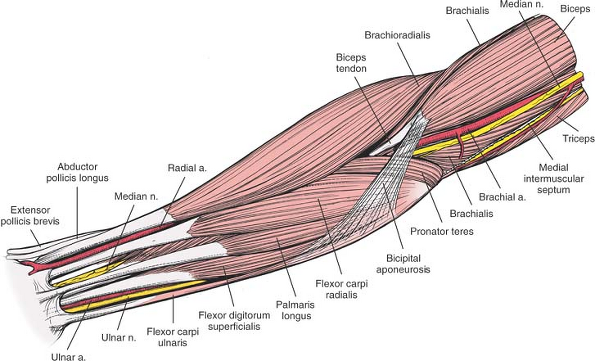 |
Figure 4-11 Superficial layer of the forearm muscles and vessels. |
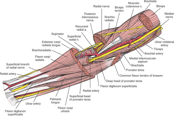 |
Figure 4-12 The superficial layer of the forearm has been resected, revealing the vessels and nerves. The median nerve pierces the gap between the two heads of the pronator teres. Note the leash of vessels of the radial artery and the recurrent radial artery. |
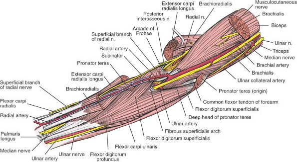 |
Figure 4-13 The middle layer of the forearm, with the superficial branch of the radial nerve. In the proximal part of the wound, the median nerve enters the undersurface of the superficialis. Flexor Carpi Radialis. Origin. Common flexor origin on medial epicondyle of humerus. Insertion. Bases of second and third metacarpals. Action. Flexor and radial deviator of wrist. Nerve supply. Median nerve. |
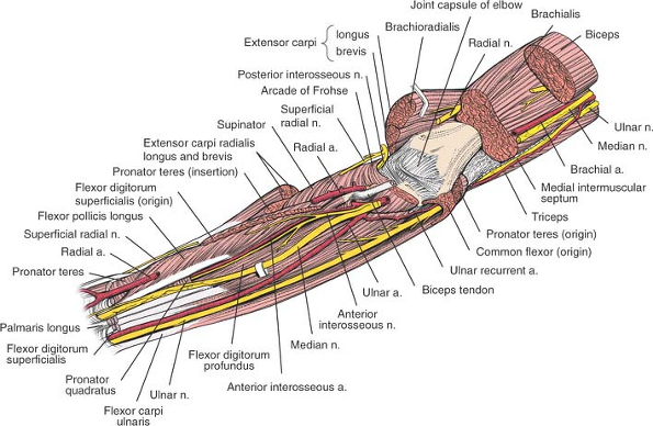 |
Figure 4-14 The deep layer of the forearm. The ulnar nerve and artery and the median nerve lie on the flexor digitorum profundus. Note the position of the anterior interosseous nerve and artery. |
Between the radial and median nerves: a dissection between the brachioradialis muscle, the most medial of the three muscles forming the “mobile wad of three” (which is supplied by the radial nerve), and the flexor carpi radialis and pronator teres muscles, the most lateral of the flexor-pronator group (which are supplied by the median nerve; see Fig. 4-3)
Between the median and ulnar nerves: a dissection between the flexor carpi ulnaris muscle (which is supplied by the ulnar nerve) and the flexor digitorum superficialis muscle, the most medial of the flexor muscles (which is supplied by the median nerve; see Fig. 5-29)
Between the ulnar and posterior interosseous nerves: a dissection between the flexor carpi ulnaris muscle (which is supplied by the ulnar nerve) and the extensor carpi ulnaris muscle (which is supplied by the posterior interosseous nerve; see Fig. 4-21)
The first of these planes is used in the anterior approach to the radius, the second exposes the ulnar nerve in the forearm, and the third is used for exposure of the ulna.
Nerves and Vessels
The neurovascular architecture of the anterior aspect of the forearm is relatively simple: the forearm is “framed” by its nerves. The superficial radial nerve runs down the radial aspect of the forearm, with the radial artery lying on its medial side in the distal half of the forearm (see Fig. 4-13). The ulnar nerve runs down the ulnar side of the forearm, with the ulnar artery lying on its lateral side in the distal half of the forearm. The median nerve runs down the middle of the forearm (see Fig. 4-14).
Both the radial and the ulnar arteries are arteries of transit in the forearm; they both are branches of the brachial artery. Because the brachial artery lies in the middle of the anterior aspect of the elbow, with the median nerve lying on its medial side, the ulnar artery and median nerve must cross in the upper forearm, with the nerve superficial to the artery; this crossing occurs at the level of the musculotendinous region of the pronator teres muscle (see Fig. 4-13). The anterior interosseous nerve (which is a branch of the median nerve) and the anterior interosseous artery (which is a branch of the common interosseous artery, which itself is a branch of the ulnar artery) also run down the middle of the forearm, but deeper than the median nerve (see Fig. 4-14).
Incision
Because the incision runs transversely across the lines of cleavage in the forearm, the resultant scar may be broad. Making the incision as a series of gentle curves brings the skin incision closer to the lines of cleavage in the forearm. Such an incision has the effect of reducing tension on the subsequent skin repair.
Superficial Surgical Dissection and Its Dangers
Muscles
Superficial surgical dissection opens the plane between the mobile wad of three muscles (the brachioradialis, extensor carpi radialis longus, and extensor carpi radialis brevis) and the pronator teres muscle proximally and flexor carpi radialis muscle distally (see Fig. 4-11).
The mobile wad of three muscles, on the radial side of the forearm, is supplied by the radial nerve. All three muscles take some of their origin from the common extensor origin on the lateral epicondyle of the humerus (see Fig. 4-13).
The brachioradialis pronates the forearm when it is supinated and supinates it when it is pronated. Therefore, it may act as a deforming force in distal radial fractures if the forearm is immobilized in either full pronation or full supination after reduction of the fracture. Its action is one reason for immobilizing distal radial fractures with the forearm in the neutral position.
The brachioradialis is the only muscle in the body to take origin from the distal end of one bone and insert onto the distal end of another (Fig. 4-15; see Fig. 4-11).
During recovery from high radial nerve palsy, the extensor carpi radialis longus is one of the first muscles to be reinnervated. If the patient recovering from a high radial nerve palsy is asked to extend the wrist, the muscle extends with radial deviation, because the balancing muscle, the extensor carpi ulnaris, is supplied farther distally by a branch from the posterior interosseous nerve. Reinnervation of the brachioradialis, however, probably is the best way to diagnose both clinically and electrically (by electromyographic studies) a recovering high radial nerve palsy (see Figs. 4-12 and 4-24).
The extensor carpi radialis brevis muscle is a wrist extensor that deviates the wrist neither toward the radius nor toward the ulna. It may be involved in tennis elbow–lateral epicondylitis.
Nerves and Vessels
Palsies of the posterior interosseous nerve caused by compression of the nerve by the tendinous origin of
the extensor carpi radialis brevis muscle have been described.
P.159
P.160
the extensor carpi radialis brevis muscle have been described.
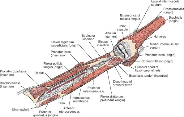 |
Figure 4-15 The origins and insertions of the muscles of the forearm. Note the anterior interosseous artery lying on the interosseous membrane. Brachioradialis. Origin. Upper two thirds of lateral supracondylar ridge of humerus. Insertion. Styloid process of radius. Action. Flexor of elbow. Pronator and supinator of forearm. Nerve supply. Radial nerve. Flexor Digitorum Superficialis. Origin. Medial epicondyle of humerus, medial ligament of elbow, medial border of coronoid process of ulna, fibrous arch connecting coronoid process of ulna with anterior oblique line of radius. Insertion. Volar aspect of middle phalanges of fingers. Action. Flexor of proximal interphalangeal joints, metacarpophalangeal joints, and wrist joint. Nerve supply. Median nerve.
Related posts:Stay updated, free articles. Join our Telegram channel
Full access? Get Clinical Tree


|
