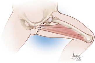
Fig. 1
Origins of the hip adductor muscles (Reprinted with permission, Cleveland Clinic Center for Medical Art & Photography © 2013. All rights reserved)
Numerous nerves and other vascular structures are at risk in dissection to perform tendon releases about the hip joint. Risk for their injury during dissection has been well documented [1]. The femoral nerve , artery, and vein lie rather superficially in the interval between the sartorius and the adductor longus/pectineus. The lateral femoral cutaneous nerve runs deep to the inguinal ligament at a point 2–3 cm medial to the ASIS. The two main branches of the obturator nerve surround the adductor brevis muscle in the groin. The anterior branch runs along the anterior portion of the adductor brevis, while the posterior branch runs posteriorly.
Surgical Management
Decisions to intervene surgically are patient specific, taking into consideration goals of treatment and function. In some cases, tendon releases about the hip joints are undertaken to avoid hip subluxation or dislocation, such as a young child with progressive subluxation. In other instances, tendon releases are performed to aid in ambulation. Some releases, such as complete psoas release from the lesser trochanter, are contraindicated in ambulatory patients. Regardless of the surgical indication, a thorough physical examination is required preoperatively to verify the character and extent of adduction or flexion contracture at the hip joint.
Routine preoperative planning usually involves supine AP radiographs (neutral/abduction), with standing radiographs obtained if patient function allows. In ambulatory patients, preoperative gait analysis can be extremely beneficial as well, giving insight into the pathologic portions of the patient’s gait. It is also recommended that a thorough examination under anesthesia be performed prior to incision. At this point, while the patient is fully anesthetized and spasticity is minimized, it is possible to differentiate between decreased range of hip motion that is caused by abnormal muscle tone and that which is caused by true musculotendinous contracture (i.e., Thomas test).
Open Adductor Lengthening and Psoas Release
The patient is placed in a supine position, with the operative leg isolated so that the hip can be ranged freely. A 2–3 cm transverse incision is made in the groin crease directly overlying the adductor longus tendon (Fig. 2). This is made 1 cm distal to the groin crease. After isolating the adductor longus tendon through careful dissection, a right-angle clamp is passed around the tendon, separating it from the underlying adductor brevis muscle. Using electrocautery, the tendon is divided at the most proximal point possible. The adductor brevis muscle can then be isolated and divided in a similar manner. Care should be taken to avoid the anterior branch of the obturator nerve, which runs along the anterior portion of the adductor brevis, and the posterior branch of the obturator nerve, which lies posterior to the adductor brevis (Fig. 3). If further release is indicated, the gracilis can be divided. This muscle is found posterior to the adductor brevis, and it tends to be much smaller and flatter in nature. If psoas release is desired, the pectineus can be identified at this point. Using cautious blunt dissection, the interval between the pectineus and neurovascular bundle is developed, revealing the underlying psoas tendon. Exposure of the psoas tendon involves dissection around the medial femoral circumflex artery (which is often ligated) and removal of a fatty collection that invariably surrounds the tendon. The psoas tendon can then be released from the lesser trochanter [2]. In addition, a musculotendinous recession can be carried out from this approach, as a full release is not recommended in an ambulatory child [3, 4] (Fig. 4).


Fig. 2


Transverse incision for open procedure (Reprinted with permission, Cleveland Clinic Center for Medical Art & Photography © 2013. All rights reserved)
Stay updated, free articles. Join our Telegram channel

Full access? Get Clinical Tree


