Fig. 1
(a) Prone position of a patient with a complete proximal hamstring rupture of the left lower extremity. (b) Prone position of a patient with a complete proximal hamstring rupture of the left lower extremity demonstrating a “popeye deformity”
Indications for Surgery
Surgical management of proximal hamstring ruptures is indicated for two- or three-tendon ruptures with greater than 1–2 cm of retraction in active, healthy individuals. With only one tendon involved or even multiple tendon involvement without significant retraction, nonoperative management may be highly successful [7]. For patients with significant comorbidities, or without a desire to return to athletic activities, nonsurgical treatment is considered. Although less successful and predictable, chronic repair or reconstruction may also be warranted secondary to pain, weakness, and loss of function after failed conservative treatment or delay in diagnosis [9, 10]. In the chronic repair or reconstruction setting, there is a possibility for increased surgical difficulty due to adhesions around the sciatic nerve, preventing sufficient tendon mobilization and warranting reconstruction with allograft [11].
Surgical Anatomy
The hamstring musculature is composed of the semimembranosus , semitendinosus , and long and short heads of the biceps femoris . The “anatomic footprint” of the proximal hamstring origin has a common semitendinosus/long head of the biceps femoris origin and separate semimembranosus origin. The semitendinosus and biceps femoris long head tendons originate on an oval-shaped, more superficial and medial area that is longer than wide (2.8 cm proximal to distal; 1.7 cm medial to lateral) [12]. The semimembranosus origin is crescent shaped, deeper, and lateral to the semitendinosus/long head of the biceps origin (3.1 cm proximal to distal; 1.1 cm medial to lateral) [12]. The mean distance from the most proximal aspect of the semitendinosus and biceps origin to the inferior margin of gluteus maximus is 6.3 cm [12]. The mean distance from the inferior gluteal nerve and artery to the inferior margin of gluteus maximus is 5.0 cm [12]. The mean distance from the sciatic nerve (lateral to the tendinous origin, entering surgical field under piriformis) to the lateral-most margin of the ischial tuberosity is 1.2 cm [12]. The posterior femoral cutaneous nerve also enters the surgical field from under the piriformis but then travels deep to the gluteus maximus and then deep to the gluteal fascia and fascia lata down the posterior thigh.
Surgical Technique
General endotracheal anesthesia with muscle relaxation is commenced, and the patient is then positioned prone on a well-padded operative table (Fig. 2a). The table is then broken at the pelvis to allow for hip flexion and forward tilting of the pelvis to rotate the ischial tuberosity into the field more clearly (Fig. 2b). Alternatively two large rolls can be used with one placed under the chest and the other under the anterior pelvis. A limb support device is necessary for sterile limb preparation and draping (Fig. 2c). Either a longitudinal (proximal to distal) or transverse (medial to lateral) incision may be used. A transverse incision in the gluteal fold/crease often provides sufficient exposure and cosmesis (Fig. 3). Even a significantly retracted (up to 5–7 cm) tendon may be adequately visualized and mobilized because of excellent skin mobility in this area. Subdermal and subcutaneous infiltration of local anesthetic can be performed prior to skin incision. The skin incision is made and sharp dissection carried down through subcutaneous tissue to identify the gluteal fascia. A transverse incision is made in the fascia with care taken to avoid the posterior femoral cutaneous nerve and its branches. A blunt retractor is placed at the inferior margin of gluteus maximus and retracted superiorly to expose the hamstring fascia. One should avoid over-retraction of the gluteus maximus to avoid inferior gluteal nerve injury. The deep hamstring fascia is incised longitudinally and a large hematoma or seroma depending on timing of surgery is often expressed and evacuated. Avoid lateral placement of this fascial exposure as the sciatic nerve is lateral to the tendinous origin. The ischium should now be visible, but deep in the surgical field (a headlamp can be used for optimal visualization). Given that the most proximal aspect of the origin is 6.3 cm from the inferior margin of the unretracted gluteus maximus, one must retract the gluteus maximus in order to provide adequate exposure and allow for anatomic reattachment of the hamstrings.
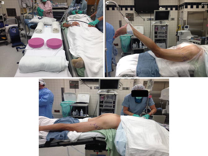
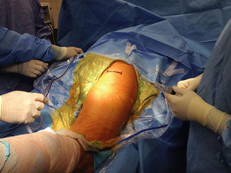
Fig. 2
(a) Operating room table prepared to pad the right and left chest walls and the right and left knees and a padded roll to elevate the ankles and flex the knees. (b) Break in the operating room table at the pelvis to allow the hips to flex and rotate the pelvis to bring the ischial tuberosity into clearer view in the operative field. (c) Lower extremity limb supporter “candy cane” to allow for sterile preparation and draping of the leg
Fig. 3
Transverse skin incision at gluteal fold/crease marked for right hip in prone position
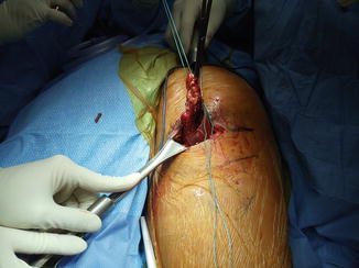
After adequate deep exposure, the retracted tendon is identified and grasped with an Allis clamp. Tagging stay sutures placed in the proximal tendon edge can aid in exposure (Fig. 4). Mobility is ensured by advancement to its anatomic footprint. Adhesiolysis is carefully performed and can be assisted with Loupe and/or microscopic magnification as needed to avoid sciatic nerve injury. The sciatic nerve should be repeatedly visualized or palpated throughout the case to avoid iatrogenic injury. The ischial footprint is next prepared with a rongeur to a light bleeding surface (Fig. 5). Double-loaded suture (nonabsorbable) anchors are then placed (usually two or three depending on patient’s anatomy and tear pattern) (Fig. 6). The surgeon should assure that the most lateral anchor is proximo-lateral enough to re-create the anatomy of the footprint. Only one limb of each suture is placed in a Krackow or locking pattern to allow for a “pulley technique .” Mark the post limb of all sutures and pull the post limbs to reduce the torn proximal tendon to the anatomic footprint (Fig. 5). When final knot tying is performed, place the hip in neutral extension and the knee flexed to 90° to provide minimal tension on the repair, if necessary. Knots are securely tied. Medial knot placement precludes contact with the sciatic nerve (lateral).
 Athletic Populations of Interest in Hip Arthroscopy and Hip Preservation Surgery
Nerve Injuries Around the Hip
Selective and Image Guided Injections Around the Hip and Pelvis
Surgical Technique: Resection Arthroplasty
Femoral Head Fractures
Surgical Technique: Osteochondral Autograft Transfer and Osteochondral Allograft Transplant for Preservation of the Femoral Head and Acetabulum
Athletic Populations of Interest in Hip Arthroscopy and Hip Preservation Surgery
Nerve Injuries Around the Hip
Selective and Image Guided Injections Around the Hip and Pelvis
Surgical Technique: Resection Arthroplasty
Femoral Head Fractures
Surgical Technique: Osteochondral Autograft Transfer and Osteochondral Allograft Transplant for Preservation of the Femoral Head and Acetabulum


Related posts:
Athletic Populations of Interest in Hip Arthroscopy and Hip Preservation Surgery
Nerve Injuries Around the Hip
Selective and Image Guided Injections Around the Hip and Pelvis
Surgical Technique: Resection Arthroplasty
Femoral Head Fractures
Surgical Technique: Osteochondral Autograft Transfer and Osteochondral Allograft Transplant for Preservation of the Femoral Head and Acetabulum
Stay updated, free articles. Join our Telegram channel
Full access? Get Clinical Tree
