Fig. 1
Observation of the skin folds on the patient’s legs will often demonstrate asymmetry of the skin folds
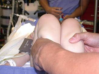
Fig. 2
Examination of the patient shows a positive Galeazzi sign with a difference in knee height. After further investigation, it was confirmed that the patient had a dislocated left hip
Table 1
Graf classificationa
Graf hip type | Descriptive term | α angle (°) | β angle (°) | Age (years) | Recommended treatment | Equivalent radiographic description |
|---|---|---|---|---|---|---|
I a | Normal | ≥60 | >70 | Any | No | Concentrically reduced |
I b | Normal | ≥60 | <70 | Any | No | Concentrically reduced |
II ab | Physiologically immature | 50–59 | nac | 0–12 weeks | Depends on age and α angle | Concentrically reduced |
II b | Immature | 50–59 | nac | >12 weeks | Yes | Concentrically reduced |
II c | Dysplastic with risk of dislocation | 43–49 | <77 | Any | Yes | Centered on medial wall of acetabulum with sloping of roof |
D | Decentered | 43–49 | >77 | Any | Yes | Subluxated |
III a | Decentered | <43 | nad | Any | Yes | Dislocated |
III b | Decentered | <43 | nad | Any | Yes | Dislocated |
IV | Decentered | <43 | nad | Any | Yes | Dislocated |
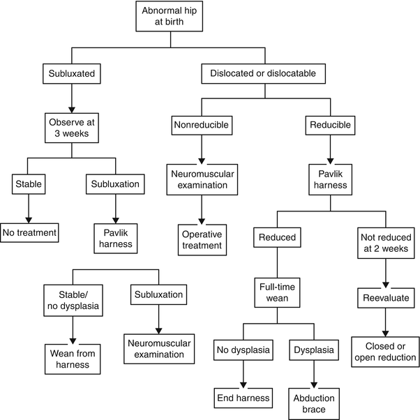
Fig. 3
Algorithm for evaluation and treatment of DDH in the newborn to 6 months of age (© 2000 American Academy of Orthopaedic Surgeons. Reprinted from the Journal of the American Academy of Orthopaedic Surgeons, volume 8(4), pp. 232–242 with permission)
If the hip has a greater degree of instability and is dislocated or dislocatable, the patient should be placed in a Pavlik harness [2, 8, 16]. Alternatively in the setting of an unreliable family or unfavorable social situation, a spica cast would be the initial choice of treatment instead of the Pavlik harness [16]. Within 2–3 weeks of stabilizing the hip in a Pavlik harness, the patient must be observed for spontaneous reduction of the hip. If the hip has been reduced, the patient should undergo a full treatment course with the Pavlik harness and periodic observation. When the hip fails to spontaneously reduce in the Pavlik harness, the patient should be considered a candidate for a closed reduction of the hip and spica casting with the option of prereduction overhead traction.
6 to 18 Months of Age
Patients between the ages of 6 and 18 months retain the ability to readily remodel the acetabulum and initially warrant nonsurgical treatment. Unfortunately, use of the harness is often precluded in children who begin to wear it after the age of 6 months. The difficulty with initiating the required immobilization in the harness has led to failure rates exceeding 50 % in older patients [18, 22]. The treatment of choice for patients 6–18 months of age would be closed reduction of the hip and spica casting with the option of an adductor tenotomy to aid the reduction (Fig. 4) [22]. Regrettably, patients with an irreducible hip or failure to have a medial dye pool of less than 7 mm on arthrography (refer to section on closed reduction and spica casting for definition of medial dye pool) following closed reduction must proceed with open reduction and spica casting [22, 23].
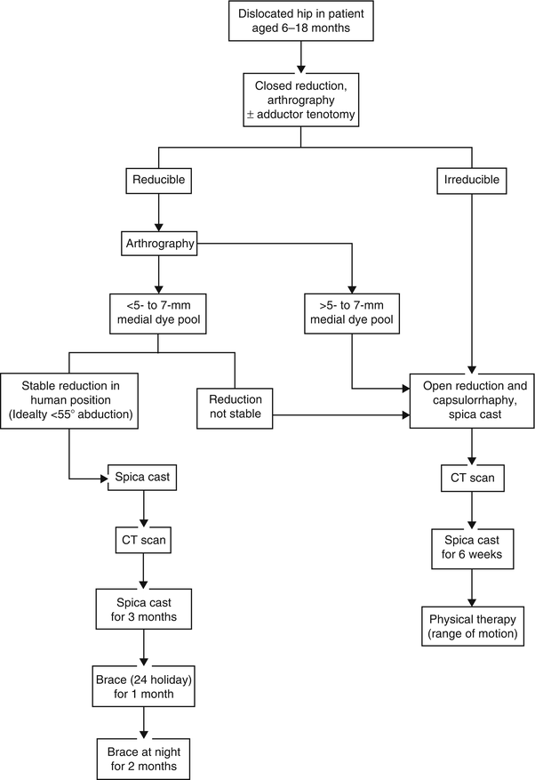
Fig. 4
Algorithm for evaluation and treatment of DDH in children aged 6–18 months of age (© 2001 American Academy of Orthopaedic Surgeons. Reprinted from the Journal of the American Academy of Orthopaedic Surgeons, volume 9(6), pp. 401–411 with permission)
18 Months of Age and Older
Older patients have a reduced capacity to remodel the acetabulum, which inhibits successful nonsurgical treatment of DDH. Despite the increased failure of closed reduction of the hip and spica casting, it is recommended that patients undergo a trail of nonsurgical treatment (Fig. 5) [22]. Because certain studies have suggested that acetabular remodeling can occur up to the age of 8 years, a trial of closed reduction and spica casting can limit the associated morbidity of an extensive open procedure [24, 25].
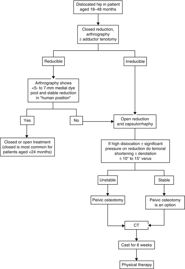
Fig. 5
Algorithm for evaluation and treatment of DDH in children aged more than 18 months (© 2001 American Academy of Orthopaedic Surgeons. Reprinted from the Journal of the American Academy of Orthopaedic Surgeons, volume 9(6), pp. 401–411 with permission)
Nonsurgical Techniques
Pavlik Harness
Since Arnold Pavlik published articles on the harness that would bear his name, other devices have been available but all have failed to offer the same flexibility of use and stability of the hip [8, 16, 26]. The original harness has evolved over the last half-century from its original construct of leather straps; however, the concept of maintaining the hips in flexion and abduction while limiting extension and adduction continues to be the foundation of today’s Pavlik harness. The use of the Pavlik harness has been accepted as the standard of treatment for DDH among infants [8, 16].
Technique
In general, the Pavlik harness consists of a chest strap, shoulder straps, and anterior and posterior stirrup straps (Fig. 6). Several manufactures offer a preassembled Pavlik harness, which all slightly differ in the application of the harness. Because the Pavlik harness can appear complicated due to the multiple straps, it is recommended to be familiar with the manufacture’s handbook and how to properly apply the specific harness. Once the Pavlik harness has been applied to the infant, the straps should be adjusted to situate the hips into the optimal position within the “safe zone” of Ramsey [16, 19]. In order to determine the “safe zone,” position the infant’s hips in 90° of flexion and passively adduct the hips noting when the femoral head displaces from the acetabulum [16, 19]. The “safe zone” is the degree difference between maximal passive abduction and when the femoral head dislocates during passive adduction [16, 19]. If the infant has a narrow “safe zone” of less than 40°, there should be consideration of an adductor tenotomy [19]. After the straps have been adjusted, no actual reduction maneuver of the hip is required. The normal movement of the infant in the harness will usually cause the hip to spontaneously reduce [2]. When infants failed to achieve hip reduction with the Pavlik harness in Ramsey’s series, he noted the most common cause of failure was inadequate hip flexion [16].
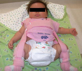
Fig. 6
Infant in the Pavlik harness demonstrates proper placement of the chest strap, shoulder straps, and anterior and posterior stirrup straps with the hips in adequate flexion and abduction
Before patients leave the office, the parents must be properly trained to remove, apply, and adjust the harness straps. It will often take significant counseling of the parents until they are comfortable with the harness. Since parents will need to be capable of adjusting the straps but likely would not be able to accurately assess the infant’s “safe zone,” typically the proper position to instruct parents is to position the hips between 100° and 110° of flexion with mild abduction [2, 17, 19]. When parents appear to have significant difficulty in the office with the harness, it is recommended to provide closer follow-up. If the parents continually have difficulty with the harness, it would be considered an indication to transition the infant to a spica cast.
Multiple variables will determine the duration of treatment and monitoring of the infant in a Pavlik harness. Once the infant has been placed in the harness, ultrasonographic evaluation should document adequate flexion and direction of the femoral head toward the triradiate cartilage [27]. The infant should wear the harness for at least 23 h a day until clinical and ultrasonographic examinations are within normal limits [8, 28]. Infants with a dislocated hip should be reevaluated every 1–2 weeks to observe for reduction of the hip, while some recommend that the infants with adequate parental support do not require further evaluation for 3–4 weeks [8, 17, 19]. If the hip remains dislocated or demonstrates no improvement in the degree of dysplasia on ultrasound after 3–4 weeks, the harness should be discontinued to prevent Pavlik harness disease and the infant is considered a candidate for closed reduction and spica casting [8, 17].
After successful reduction of the hip, the infant only needs follow-up every 3–4 weeks for reevaluation. In general, the duration of treatment in the Pavlik harness is a minimum of 3 months for infants 3 months of age or younger, but older children should remain in the harness for approximately double their age [8]. It is not necessary for the harness to be worn 23 h a day during the entire duration of treatment. Beginning at the midpoint of treatment, the hours spent in the harness can gradually be weaned [16]. Prior to weaning the harness, the parents should take the patient out of the harness the night before the office visit [16]. If the ultrasonographic evaluation the following day is consistent with a stable hip, the infant can increase to 4 hours per day spent out of the harness for the first third of the remaining treatment period, then 8 hours per day for the second third of the treatment period, and then 12 hours per day for the last third of the duration of treatment. At the end of the weaning process, check for residual acetabular dysplasia with the use of ultrasonography. If present, continue to wear the harness 12 h per day until the patient has radiographic resolution of the dysplasia [16].
Several contraindications exist in the use of a Pavlik harness to treat DDH. Because reduction of the hip is reliant on normal movements of the infant, any delay in motor skills or major muscle imbalance would preclude the use of the Pavlik harness [2, 8]. After reduction of the hip, the Pavlik harness along with the soft tissue helps to maintain the reduction. Therefore, infants with ligamentous laxity as in Ehlers-Danlos syndrome would not benefit from the harness [8]. As previously stated, the infant’s age and family situation would be limiting factors and an indication for another treatment option [2, 28]. Although not contraindications, historically cited risk factors for failure of the Pavlik harness are an absent Ortolani sign at the initial evaluation, bilateral dislocation, and age of greater than 7 weeks before initiation of treatment [29]. More recently, a systematic review on the Pavlik harness identified the radiographic distance from the midpoint of the proximal metaphyseal border of the femur to Hilgenreiner’s line on the initial radiograph as predictive of success and failure of the Pavlik harness [30]. The literature has demonstrated that a value greater than 8 mm is associated with a reduced risk of AVN and a value greater than 6 mm was indicative of a satisfactory outcome [31–33].
Closed Reduction and Spica Casting
When children fail or exhibit a contraindication to the Pavlik harness, the remaining alternative to surgical treatment is closed reduction and spica casting . Historically, closed reduction of the hip was preceded by the use of longitudinal or overhead traction. More recently, the use of traction has become controversial due to complications of skin breakdown and the belief that it does not aid in the reduction [22]. Advocates for prereduction traction believe the gentle stretching of the soft tissue structures improves the success of closed reduction and reduces the risk of osteonecrosis. However, the current literature has conflicting reports on the use of prereduction traction [22, 34, 35].
Technique
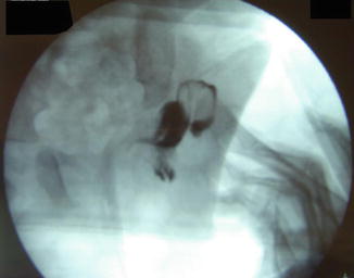
Closed reduction of the hip should be performed under general anesthesia in the operating room to provide adequate muscle paralysis. The reduction maneuver involves longitudinal traction, flexion, and abduction of the hip, all while applying posterior pressure on the greater trochanter [16]. Frequently an adductor tenotomy is necessary via an open or percutaneous technique, which relieves one of the opposing forces and widens the “safe zone.” After reduction of the hip, intraoperative arthrography will confirm a concentric reduction of the femoral head by demonstrating a collection of dye in the space between the femoral head and medial border of the acetabulum of less than 5–7 mm (Figs. 7 and 8) [23]. The previously described collection of dye is often referred to as the “medial dye pool.” If the medial dye pool measures greater than 7 mm, it is an indication to proceed with an open reduction [23].
 Neuromuscular Hip Disorders: Focus on Cerebral Palsy
 Femoral Deformities: Varus, Valgus, Retroversion, and Anteversion
 Surgical Technique: Arthroscopic Labral Management
 Surgical Technique: Endoscopic Gluteus Medius Repair
 Subspine Impingement and Surgical Technique
 Atraumatic Instability and Surgical Technique


Related posts:
Neuromuscular Hip Disorders: Focus on Cerebral Palsy
Femoral Deformities: Varus, Valgus, Retroversion, and Anteversion
Surgical Technique: Arthroscopic Labral Management
Surgical Technique: Endoscopic Gluteus Medius Repair
Subspine Impingement and Surgical Technique
Atraumatic Instability and Surgical Technique
Stay updated, free articles. Join our Telegram channel
Full access? Get Clinical Tree
