Fig. 1
Lateral view of gluteus medius anatomy and insertion site (Reprinted with permission, Cleveland Clinic Center for Medical Art & Photography © 2013–2014. All rights reserved)
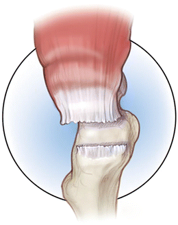
Fig. 2
Schematic depiction of gluteus medius tear (Reprinted with permission, Cleveland Clinic Center for Medical Art & Photography © 2013–2014. All rights reserved)
Gluteus medius tendon tears are typically seen in three scenarios: (1) chronic, nontraumatic tear of the anterior fibers of the gluteus medius tendon; (2) abductor tendon tears found in patients with femoral neck fractures or osteoarthritis; and (3) avulsion after total hip arthroplasty performed through an anterolateral or transgluteal approach. The most common of these scenarios is chronic, nontraumatic tears.
Several repair techniques have been described including repairs using both transosseous sutures [2, 9, 10] and suture anchors [11]. Endoscopic repair techniques include gluteal debridement or repairs, bursectomy, and iliotibial band release [1, 12–15]. Gluteus maximus muscle transfers [16–19] and vastus lateralis muscle transfers [20, 21], dermal matrix allograft augmented repair techniques [22], and Achilles tendon allograft [23] have been described as well.
Few reports have been published on the outcomes of open repairs. The limited data from small patient series mainly presented as part of surgical technique papers suggest overall good outcomes, however often requiring extensive rehabilitation and long recovery times [12, 24].
Surgical Technique
The patient is placed supine on a standard operating room table. After induction of general endotracheal anesthesia, the patient is turned to a lateral decubitus position on a well-padded peg board. All bony prominences are well padded and a pneumatic compression device is placed on the well leg. After standard preparation of the operative extremity and sterile draping, a lateral skin incision centered over the greater trochanter is used to develop the interval between subcutaneous tissue and underlying iliotibial band. The iliotibial band is split longitudinally in line with its fibers, and appropriate retractors are placed, permitting visualization of the vastus ridge and the lateral facet of the greater trochanter. This allows entry into the peritrochanteric space to assess the gluteus medius tear (Fig. 4). The hip is internally rotated, exposing the trochanteric bursa at the posterior facet. The short external rotators are assessed for integrity. The sciatic nerve is typically not visualized or exposed.
The extremity is then externally rotated revealing the anterior trochanteric facet at the former insertion site of the gluteus minimus. Residual soft tissue remnants are thoroughly debrided off the footprint of the gluteus medius and minimus using a rongeur and round burr, promoting a favorable biologic healing response (Fig. 5). The retracted gluteus medius and gluteus minimus tendons are identified and assessed for visible degenerative changes in either tendon. Suture anchors are then placed into the peripheral zones of the footprint (see Fig. 3). The number of anchors placed is based on the size for the tear. Sutures can be passed in multiple manners. Options include simple, mattress, modified Mason-Allen, or a running locking fashion throughout the periphery of the crescent-shaped tear in the gluteus medius and minimus. Suture configuration is guided by surgeon preference and tear configuration. As in the shoulder, double-row fixation or transosseous equivalent type configurations can also be used. Sutures are tied with the leg held in slight abduction (10–15°) to limit tension at the repair site and reduce the tendon onto the prepared footprint (Fig. 6). On completion of the repair, the hip is gently brought through a broad physiologic range of motion and rotation. The footprint of the gluteus medius should be intact and stable throughout this range of motion.
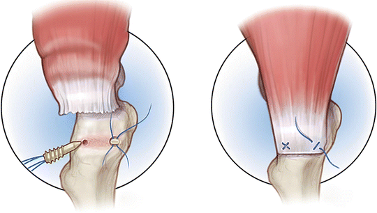
Fig. 3
Schematic depiction of gluteus medius repair with anchors (Reprinted with permission, Cleveland Clinic Center for Medical Art & Photography © 2013–2014. All rights reserved)
Rehabilitation
Postoperatively, the patient is restricted to 20-lb flat-foot weight bearing for 6 weeks with no active abduction or passive adduction. A hip abduction brace is used during this time period, with a pillow between the legs while sleeping. The patient is advanced to weight bearing as tolerated by 8 weeks and full weight bearing without assistive devices by 12 weeks. Active hip abduction is allowed after the 8-week mark and progressive strengthening employed once full range of motion attained.
Outcomes
There are several small case reports in the literature on patients treated with open surgical repair of hip abductor tears [2, 11, 13, 26–29]. Most reports show good pain relief and some improvement in function after surgical repair. Clinical follow-up and long-term outcome assessment is limited, though. Davies et al. reported a retrospective review of a case series including 22 patients (23 hips). The mean Harris hip score improved from 53 points preoperatively to 87 points at 1 year and 88 points at 5 years. The mean lower-extremity activity scale score improved from 6.7 points preoperatively to 8.9 points at 1 year and 8.8 points at 5 years. There was no significant difference in the degree of clinical improvement in relation to the severity of the tear. However, three patients had poor results and were part of the group with the largest tears. Sixteen of nineteen patients were satisfied with their outcome and willing to undergo the procedure again. Davies et al. reported on 16 patients who underwent open surgical repair [26]. There were 4 re-ruptures, 3 of which were revised and 1 deep infection requiring debridement. The remaining 11 patients had statistically significant improvements in hip symptoms. The mean change in visual analogue score was reported as 5 out of 10 (p = 0.0024). The mean change of Oxford hip score was 20.5 (p = 0.00085). The mean improvement in SF-36 PCS was 8.5 (p = 0.0020) and MCS 13.7 (P = 0.134). Six patients with preoperative Trendelenburg gait had normal gait 1 year following surgery. They concluded that surgical repair is overall successful for reduction of pain and improvement of function, but that there is a relatively high failure rate in chronic tears. Walsh et al. reported results of open surgical repair in 72 patients with a minimum follow-up of 1 year [28]. Improvement in both function and pain over time was seen in 95 % of their patients. Voos et al. reported good pain relief 2 years after arthroscopic repair in ten patients with low-grade tears [13]. Open repair was recommended for larger tears.
Related posts:
 Athletic Populations of Interest in Hip Arthroscopy and Hip Preservation Surgery
Nerve Injuries Around the Hip
Selective and Image Guided Injections Around the Hip and Pelvis
Surgical Technique: Resection Arthroplasty
Surgical Technique: Arthroscopic Treatment of Chronic Slipped Capital Femoral Epiphysis
Smith-Petersen Approach to the Hip
Athletic Populations of Interest in Hip Arthroscopy and Hip Preservation Surgery
Nerve Injuries Around the Hip
Selective and Image Guided Injections Around the Hip and Pelvis
Surgical Technique: Resection Arthroplasty
Surgical Technique: Arthroscopic Treatment of Chronic Slipped Capital Femoral Epiphysis
Smith-Petersen Approach to the Hip
Stay updated, free articles. Join our Telegram channel

Full access? Get Clinical Tree


