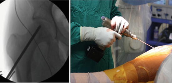
Fig. 1
A guide wire is passed through the lateral femoral cortex into the necrotic area of the femoral head. The starting point should not be distal to the lesser trochanter as this may increase the risk of subtrochanteric fracture

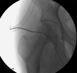
Fig. 2
A cannulated drill is introduced over the guide wire into the subchondral bone. This is repeated in several directions to fully decompress the necrotic lesion

Fig. 3
Anteroposterior intraoperative view of the hip after the core has been filled with autologous bone graft followed by calcium phosphate bone void filler (Norian)
The rehabilitation protocol includes non-weight bearing with crutches for the first 6 weeks to minimize the risk of femoral neck fracture. The patient will begin supervised physical therapy immediately postoperatively to maintain hip range of motion. At 6 weeks after surgery, the patient is allowed to progressively weight bear until full weight bearing is achieved.
Summary
AVN often afflicts a younger demographic, making the prospect of undergoing THA at this age a difficult decision. Hip prostheses in this demographic often undergo greater wear than in older patients, which is important as the average survivorship of a total hip implant is inversely proportional to activity level [30], setting the patient up for several revision surgeries over his lifespan. Moreover, in a more athletic demographic, hip resurfacing and THA often signify the end of a competitive athletic career; therefore these procedures are typically deferred to as latter options [31]. In appropriately selected patients, core decompression with bone graft is a viable and reliable option to delay the need for arthroplasty.
References
1.
Mont MA, Hungerford DS. Non-traumatic avascular necrosis of the femoral head. J Bone Joint Surg Am. 1995;77(3):459–74.PubMed
2.
3.
Etienne G, Mont MA, Ragland PS. The diagnosis and treatment of nontraumatic osteonecrosis of the femoral head. Instr Course Lect. 2004;53:67–85.PubMed
4.
5.
Sen RK. Management of avascular necrosis of femoral head at pre-collapse stage. Indian J Orthop. 2009;43(1):6–16.PubMedCentralPubMedCrossRef
6.
Steinberg M. Diagnostic imaging and role of stage and lesion size in determining outcome in osteonecrosis of the femoral head. Tech Orthop. 2001;16:6–15.CrossRef
7.
8.
Jergesen HE, Heller M, Genant HK. Signal variability in magnetic resonance imaging of femoral head osteonecrosis. Clin Orthop Relat Res. 1990;253:137–49.PubMed
Stay updated, free articles. Join our Telegram channel

Full access? Get Clinical Tree


