Fig. 1
(a–c) (a) Line drawing showing the reflected (indirect) head of the rectus femoris spanning over the defect of the acetabulum. Note its “Y” attachment to the direct head of the rectus femoris, (b) the indirect head has been partially detached from its insertion on the ileum leaving the distal attachment intact and an interim attachment to the muscular portion of the direct head, (c) the indirect head attached to the rim of the acetabulum with suture anchors; note the distal attachment remains intact and the direct head attachment has been released. Note the side-to-side anastomosis (arrow)
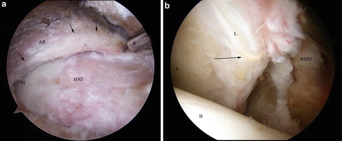
Fig. 2
(a–b) (a) View of a left hip assessing the labral quality; note the degeneration and poor tissue (arrows), acetabular rim (AR), femoral head (H), and head neck junction (HNJ); (b) remaining loop suture (arrow) around a stiff nonfunctional labrum (L), acetabulum (A)
Through an extensive capsulotomy, expose the rim of the acetabulum and the area to be grafted (Fig. 3). Identify the indirect head of the rectus femoris and release it from its superior attachment to the ilium, leaving the lateral insertion and the muscular attachments intact (Fig. 4). The idea is to create a “bucket handle” which will be sequentially attached to the acetabular rim from lateral to anterior while leaving the peripheral attachments to tension it while laying onto the rim.
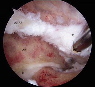
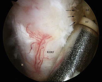
Fig. 3
View of a left hip through the anterolateral portal showing the area to be grafted after rim trimming of acetabulum; acetabular rim (AR), reflected head rectus femoris (RHRF), capsule (C)
Fig. 4
View of a right reflected head of the rectus femoris prior to detachment from the acetabular rim (arrows) by an RF wand (RF)
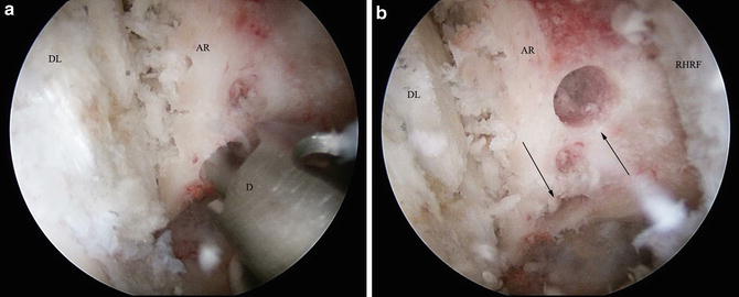
The anchor holes are predrilled usually one per zone (Fig. 5). Using any suture technique, horizontal mattress sutures are places with high-strength no-absorbable suture. Knotted or knotless anchors may be employed. As the graft is laid down and the suture is tightened forcing the graft into position, the muscular attachment may need to be partially released in sequence while still tensioning the graft (Fig. 6). Once the graft has covered the desired area, the muscular attachment is completely released to do a side-to-side anastomosis with the remaining labrum if desired.
 Athletic Populations of Interest in Hip Arthroscopy and Hip Preservation Surgery
Nerve Injuries Around the Hip
Selective and Image Guided Injections Around the Hip and Pelvis
Surgical Technique: Resection Arthroplasty
Femoral Head Fractures
Surgical Technique: Osteochondral Autograft Transfer and Osteochondral Allograft Transplant for Preservation of the Femoral Head and Acetabulum
Athletic Populations of Interest in Hip Arthroscopy and Hip Preservation Surgery
Nerve Injuries Around the Hip
Selective and Image Guided Injections Around the Hip and Pelvis
Surgical Technique: Resection Arthroplasty
Femoral Head Fractures
Surgical Technique: Osteochondral Autograft Transfer and Osteochondral Allograft Transplant for Preservation of the Femoral Head and Acetabulum


Related posts:
Athletic Populations of Interest in Hip Arthroscopy and Hip Preservation Surgery
Nerve Injuries Around the Hip
Selective and Image Guided Injections Around the Hip and Pelvis
Surgical Technique: Resection Arthroplasty
Femoral Head Fractures
Surgical Technique: Osteochondral Autograft Transfer and Osteochondral Allograft Transplant for Preservation of the Femoral Head and Acetabulum
Stay updated, free articles. Join our Telegram channel
Full access? Get Clinical Tree
