Fig. 1
Microfracture hole in the subchondral bone. A microfracture depth of 2–4 mm is made to access the marrow elements and provide a pathway for release of the underlying mesenchymal stem cells
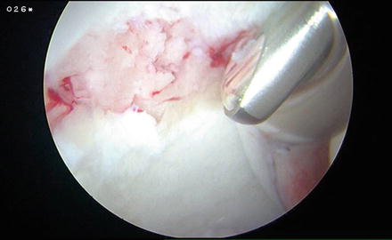
Fig. 2
The pump pressure is decreased to observe the release of blood and fat droplets from the microfracture holes
Working in the hip presents a few technical challenges that are different than those found in the knee. One of the more difficult obstacles is the ability to access different parts of the hip with an instrument and create microfracture holes that are perpendicular to the cartilage surface. Various companies are vested in the development of devices that can help surgeons overcome these obstacles.
Stryker (Mahwah, NJ) has developed a MicroFXTM OCD system with multiple curette and microfracture guide options to facilitate access to different parts of the hip. Their curettes are ring, pear, and reverse triangle shaped to help create vertical shoulders that are mechanically stable. The guides have angled necks including 0°, 45°, 70°, and 90°. The working lengths of the curettes and microfracture guides are 3.5 and 7 in. in length. Snap caps can be used with the guides and are available in 4-, 5-, 6-, and 7-mm options to allow surgeons to choose an appropriate depth of microfracture (Fig. 3). The drill has a 2-mm tip with flutes to allow for bone extraction rather than compaction. Extracting bone leaves clear channels for the efflux of marrow cells [13] (Fig. 4). The microfracture guide has a mouth that has teeth to reduce the risk of skiving (Fig. 5).
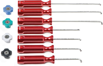
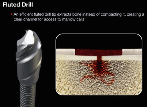
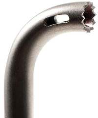
Fig. 3
Stryker snap caps (left) and microfracture guides (right). The guides have angled necks including 0°, 45°, 70°, and 90°. The working lengths of the guides are 3.5 and 7 in. in length. Snap caps can be used with the guides and are available in 4-, 5-, 6-, and 7-mm options to allow surgeons to choose an appropriate depth of microfracture (Image courtesy of Stryker, Mahwah, NJ)
Fig. 4
Stryker drill has a 2-mm tip with flutes to allow for bone extraction rather than compaction. Extracting bone leaves clear channels for the efflux of marrow cells (Image courtesy of Stryker, Mahwah, NJ)
Fig. 5
Stryker microfracture guide has a mouth that has teeth to reduce the risk of skiving (Image courtesy of Stryker, Mahwah, NJ)
Smith & Nephew (Andover, MA) has developed an XL Microfracture Pick that is added to its hip instruments tray (Fig. 6). The pick has an angle of 90° (Fig. 7). The pick has a mid-shaft strike point allowing for the 90° angle pick to be impacted with a perpendicular force into the subchondral bone (Fig. 8). This mid-shaft strike point also helps minimize the chances of skiving. The shaft is tapered and has a working length of 15.2 in., thus allowing it to be used with a slotted cannula in a standard arthroscopy portal.
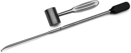
Fig. 6
 Athletic Populations of Interest in Hip Arthroscopy and Hip Preservation Surgery
Nerve Injuries Around the Hip
Selective and Image Guided Injections Around the Hip and Pelvis
Surgical Technique: Resection Arthroplasty
Femoral Head Fractures
Surgical Technique: Osteochondral Autograft Transfer and Osteochondral Allograft Transplant for Preservation of the Femoral Head and Acetabulum
Athletic Populations of Interest in Hip Arthroscopy and Hip Preservation Surgery
Nerve Injuries Around the Hip
Selective and Image Guided Injections Around the Hip and Pelvis
Surgical Technique: Resection Arthroplasty
Femoral Head Fractures
Surgical Technique: Osteochondral Autograft Transfer and Osteochondral Allograft Transplant for Preservation of the Femoral Head and Acetabulum


Smith & Nephew XL Microfracture Pick. Note the mid-shaft strike point, which allows for the 90° angle pick to be impacted with a perpendicular force into the subchondral bone (Image courtesy of Smith & Nephew, Andover, MA)
Related posts:
Athletic Populations of Interest in Hip Arthroscopy and Hip Preservation Surgery
Nerve Injuries Around the Hip
Selective and Image Guided Injections Around the Hip and Pelvis
Surgical Technique: Resection Arthroplasty
Femoral Head Fractures
Surgical Technique: Osteochondral Autograft Transfer and Osteochondral Allograft Transplant for Preservation of the Femoral Head and Acetabulum
Stay updated, free articles. Join our Telegram channel
Full access? Get Clinical Tree
