Fig. 1
Hip chondral lesions : surgical alternatives and novel technique with platelet-rich plasma and mononuclear cells concentrate. (a–c) Standard alternatives for hip chondral lesions. (a) Microfractures. (b) Thermal chondroplasty. (c) Chondral flaps resection. (d) and (e) Novel surgical technique. The PRP clot is positioned over the microfractured area and mononuclear cells concentrate is instilled under it. (f) and (g) dGEMRIC images at 6 months postop of the same patient, in which a homogenous captation of gadolinium is observed, meaning the restoration of glycosaminoglican content
Excellent results have been obtained with this technique, confirming the restoration of glycosaminoglycan concentration by MRI metabolic-type dGEMRIC (delayed gadolinium-enhanced magnetic resonance imaging of cartilage) (Fig. 1f, g).
Surgical Technique
After rim trimming and labrum refixation, cartilage assessment is made. If chondral lesion exists, the harvesting of 15 cc of bone marrow is made and centrifugated. obtaining 2–4 cc of autologous bone marrow – mesenchymal stem cell concentrate (average 14 millions of nucleated cells/cc3). At the same time, 50 cc of peripheral blood is taken and centrifugated twice, in order to obtain 4 cc of PRP (6–9×), ready to be activated with autologous thrombin. Treatment of chondral lesion is made as described by Steadman in the knee, with debridement of all remaining unstable cartilage, followed by the removal of the calcified plate. After preparation of the bed, multiple holes in the exposed subchondral bone plate are made, leaving about 3–4 mm between each. Once microfracture is complete, traction is released and femoral osteoplasty is completed, obtaining a free range of motion with no abnormal contact between acetabular rim and femoral neck-head junction. At the end of the procedure, traction is reinstalled and the final part of the procedure is performed. After activation of platelet-rich plasma and clot formation, a slotted cannula is inserted via the anterior portal. Platelet-rich plasma clot is inserted through the cannula and positioned over the microfractured area. A 21-gauge trocar is then inserted passing through previously located clot and autologous bone marrow – mesenchymal stem cell concentrate is instilled under PRP clot. Traction is then released and the procedure is finished.
Rehabilitation protocol: Passive motion device is maintained for 8 h. Two crutches with partial weight bearing are indicated for 6–8 weeks. Progressive physical activities are allowed.
Preliminary Results
At the time, 13 patients with chondral lesion of the hip had been treated with microfractures and autologous bone marrow – mesenchymal stem cell concentrate transplanted on a platelet-rich plasma clot. All patients’ symptoms improved over the follow-up period of 8 months (4–12 months). Average Hip Outcome, Vail Hip, and Modified Harris Hip scores for all patients showed significant improvement at 3 and 6 months. dGEMRIC of 4 patients at 6 months postoperatively revealed complete defect fill and complete surface congruity with native cartilage.
Intra-articular Injections of Expanded Mesenchymal Stem Cells
Osteoarthritis (OA) is the most common type of arthritis and the leading cause of disability in the United States [29]. Several systemic treatments, mostly symptom-modifying rather than disease-modifying agents, are available for OA [30]. However, there is a real need for effective and safe disease-modifying OA therapies that can not only effectively treat those with established OA but also possibly delay or prevent progression in those with early OA [31]. As it was mentioned before in focal chondral lesions, mesenchymal stem cells represent a valid alternative for treatment, but multiple chondral lesions or established osteoarthritis are not suitable for focal treatment.
Adult mesenchymal stem cells were originally believed to only differentiate into tissue-specific cells. However, these cells have two major properties that could explain some of the results seen with the intra-articular injections of expanded mesenchymal stem cells, and these are homing and response to specific signals. Homing is a particular property of these cells, meaning that they respond to systemic stimuli and “travel to the place that needs repair.” The homing effect has been demonstrated in several animal studies, using labeled mesenchymal stem cells administered via systemic intravascular route or by direct local implantation, showing the presence of the marker at the injury site [32]. Mesenchymal stem cells have the ability to differentiate into a different tissue in response to specific signals released by the injury site, such as chondrogenic lineage in an osteoarthritic joint [33].
Hip extensive damage or mild OA is usually treated with local infiltrations, symptom-modifying treatments, pain killers, and finally a total hip replacement, but an increasing number of active patients seek for a non-arthroplasty treatment, and stem cells may present as an alternative to this group of patients. Intra-articular injections of expanded mesenchymal stem cells have not been described in the hip joint; however, there are some animal and clinical studies in other joints. Mokbel et al. labeled autologous adult stem cells suspended in hyaluronic acid were injected intra-articularly into carpal joints in an experimental arthritis induced by intra-articular (IA) amphotericin-B in donkeys [33]. Significant improvement was noted in clinical and radiographic OA, and significantly lesser histopathological changes of OA were seen in carpal joints that received stem cells compared to control contralateral joints. Importantly, injected stem cells were incorporated into the articular cartilage of the injected joint, as evident by their integration in the surface of the cartilage and also the interior of the cartilage [33]. Emadedin et al. injected expanded mesenchymal stem cells in six female patients with OA that required joint replacement. At 12 months follow-up, there was a significant decrease in mean pain, as well as improvements in joint functioning, walking distance, time to gelling, patellar crepitus, and knee flexion. Magnetic resonance images (MRI) obtained at 6 months after treatment showed an increase in cartilage thickness and extension of the repair tissue over the subchondral bone in half of the patients, in addition to a decrease in subchondral bone edema [34]. Mcllwraith et al. evaluated intra-articular injection of bone marrow-derived mesenchymal stem cells to augment healing with microfractures in horses. At 6 months, arthroscopic and gross evaluation confirmed a significant increase in repair tissue firmness and a trend for better overall repair tissue quality in treated joints compared to microfractures alone. Immunohistochemical analysis showed significantly greater levels of aggrecan in repair tissue treated with stem cell injection [35].
In summary, the use of intra-articular injections of expanded mesenchymal stem cells in OA has little published evidence; however, in a young active patient, it seems to be a promising non-arthroplasty treatment.
Treatment of Choice
The treatment of choice for hip diffuse chondral damage and mild osteoarthritis in an active patient seeking for a non-arthroplasty treatment is intra-articular injections of expanded mesenchymal stem cells. The surgical technique is described below.
Excellent results have been obtained with this technique, with an increase in hip functional scores (Vail-10 hip score and Harris Hip Score) and a decrease in mean pain values.
Surgical Technique
Patients were placed on an operating table in the prone position under general anesthesia. The harvesting of 15 cc of bone marrow is made and centrifugated, obtaining 2–4 cc of autologous bone marrow – mesenchymal stem cell concentrate (average 14 millions of nucleated cells/cc3) (Fig. 2). Bone marrow concentrate is then processed in a GMP laboratory, and over a 1-month period, mesenchymal stem cells are expanded to 20 × 106 cells and taken to the hospital in a portable incubator. Under fluoroscopy, cells were injected into the patients’ hips (Fig. 3).
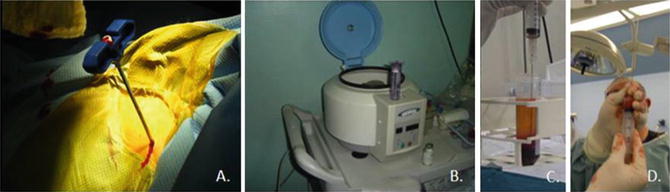
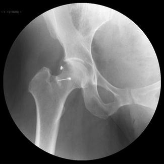
Fig. 2
Autologous mesenchymal stem cells concentrate. (a) Autologous bone marrow autograft (b) Centrifugation process with a single spin. (c) Layer separation by a density filter and identification of mononuclear cells layer. (d) Autologous mononuclear cells concentrate – final view
Fig. 3
Intra-articular injection of expanded mesenchymal stem cells . Fluoroscopic image
Preliminary Results
At the time, 7 patients with mild OA of the hip had been treated with intra-articular injections of expanded mesenchymal stem cells. All patients’ symptoms improved over the follow-up period of 10 months (8–14 months). Average Vail-10 and Modified Harris Hip scores for all patients showed significant improvement at 3 and 6 months. None of the patients has required a total hip replacement at the time.
Expanded Mesenchymal Stem Cells Seeded in a Collagen Membrane
As mentioned in Chap. 91, “Stem Cell Therapy for Hip Cartilage Lesions: General Concepts and Basic Science,” hip chondral lesions can be an elusive source of pain, and their treatment is limited to chondroplasty and debridement in partial defects and microfractures for full-thickness chondral lesions. Microfracture involves penetration of the subchondral bone to release blood and bone marrow into the defect, initiating cartilage repair. This technique has produced good clinical results in defects <2 cm2. For larger lesions, bone marrow concentrate in a PRP clot seems to be a good alternative. Other treatment options include autologous chondrocyte implantation (ACI), matrix-induced ACI (MACI), autologous matrix-induced chondrogenesis (AMIC), and membrane seeded with expanded mesenchymal stem cells.
ACI has been used increasingly for the repair of larger chondral lesions in the knee. For hip chondral lesion management, only two reports were found. Fontana et al. described a case control study in 30 patients with hip chondral lesions , 15 treated with ACI and 15 with debridement alone. At 74 months follow-up, Harris Hip Score was significantly better in ACI group compared to the debridement group [36]. Akimau et al. described a case of severe chondrolysis and osteonecrosis of the femoral head after a severe fracture dislocation in a 31-year-old man. Twenty-one months after the injury, they performed a MACI technique. At 1-year follow-up, the subjective hip score and range of motion had increased compared to preoperative values. At 15 months follow-up, biopsy demonstrated a 2 mm thickness cartilage, well populated with viable cells and integrated with the underlying bone [37].
Related posts:
 Athletic Populations of Interest in Hip Arthroscopy and Hip Preservation Surgery
Nerve Injuries Around the Hip
Selective and Image Guided Injections Around the Hip and Pelvis
Complications with Hip Arthroscopy and Open Hip Surgery
Surgical Technique: Arthroscopic Treatment of Chronic Slipped Capital Femoral Epiphysis
Smith-Petersen Approach to the Hip
Athletic Populations of Interest in Hip Arthroscopy and Hip Preservation Surgery
Nerve Injuries Around the Hip
Selective and Image Guided Injections Around the Hip and Pelvis
Complications with Hip Arthroscopy and Open Hip Surgery
Surgical Technique: Arthroscopic Treatment of Chronic Slipped Capital Femoral Epiphysis
Smith-Petersen Approach to the Hip
Stay updated, free articles. Join our Telegram channel

Full access? Get Clinical Tree


