Total hip arthroplasty
Open reduction internal fixation of the femoral neck/head
Surgical dislocation of the hip
Open reduction of congenitally dislocated hip
Labral repair
Femoral or acetabular osteo-/chondroplasty
Irrigation and debridement of septic hip
Biopsy
Excision of ectopic bone
Hip resurfacing
Osteotomies for the treatment of developmental hip dysplasia
Decompression and bone grafting for avascular necrosis of the femoral head
Hip arthrodesis
The true Hueter or Smith-Petersen approach utilizes an 8–10 cm incision from 2 to 3 cm lateral to the anterior superior iliac spine (ASIS) toward the lateral patella. The superficial internervous plane is between the sartorius (femoral nerve) and tensor fascia lata (TFL, superior gluteal nerve). The deep internervous plane lies between the rectus femoris (RF, femoral nerve) and the gluteus medius and minimus (GMed and GMin, superior gluteal nerve). Developing the interval between the TFL and sartorius directly places the lateral femoral cutaneous nerve (LFCN) at risk. The LFCN typically crosses under the inguinal ligament medial to the ASIS and courses over the TFL 1.5–5 cm distal to the ASIS [2]. However, many anatomical variants have been described and impairment of the LFCN after anterior approach for total hip arthroplasty has been reported to be up to 14.8 % [3, 4]. Matta [5] has described a modification of the Hueter approach with a more lateral skin incision, which utilizes the same superficial and deep internervous planes but protects against injury to the LFCN.
The primary goal of this chapter is to describe, in detail, the surgical technique of this modified Hueter/Smith-Petersen anterior approach to the hip.
Surgical Technique
The patient can be placed supine on a standard or orthopedic table depending on the procedure and surgeon preference. The surgical side of the patient may also be elevated or the table tilted in a semi-lateral position, which can help in obtaining a lateral fluoroscopic image of the hip. The area between the xiphoid cranially and mid-femur as well as the pubic symphysis to the posterior buttock should be prepped to allow for possible extension of the exposure (Fig. 1a). Smaller areas of interest may be draped for specific procedures (Fig. 1b).
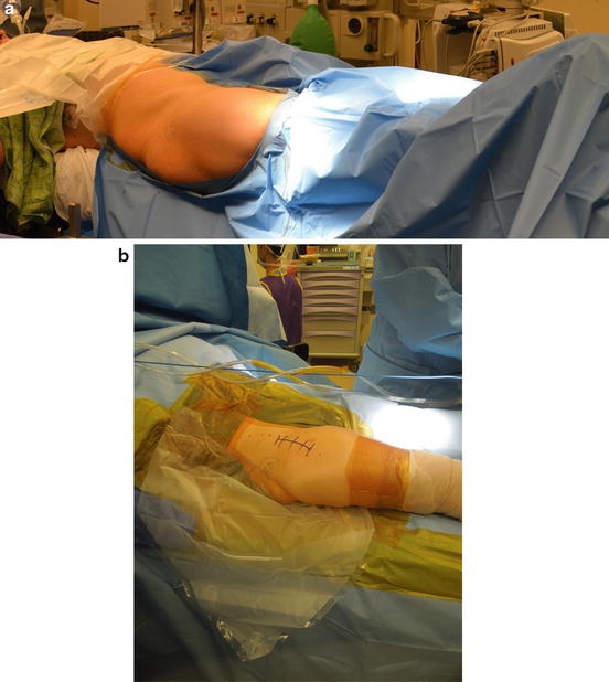
Fig. 1
(a) Initial positioning for the anterior approach on the orthopedic table. (b) Final field preparation and draping with leg free on radiolucent table
Landmarks and Incision
The iliac crest, greater trochanter, and the ASIS are identified and marked. The incision starts approximately 2 cm lateral and 1 cm distal to the ASIS, depending on the size of the patient. Continue the incision distally and posteriorly for 8–10 cm toward the anterior border of the femur in a direction parallel to the fibers of the TFL. The distal end of the incision typically lies 2–3 cm anterior to the greater trochanter. Sharply traverse the subcutaneous tissue with a scalpel and coagulate any small superficial vessels. Conversely, a transverse incision can be made in the inguinal fold [6], which can improve cosmesis. Dissection will continue down to muscular fascia. With a transverse incision, proximal and distal cutaneous flaps are developed. The translucent fascia overlying the fibers of the TFL is identified (Fig. 2).
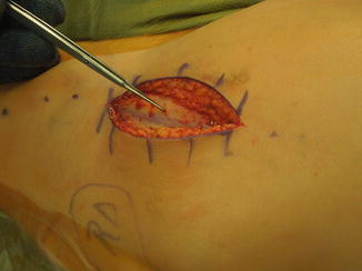
Fig. 2
Translucent fascia overlying tensor fascia lata (TFL) muscle
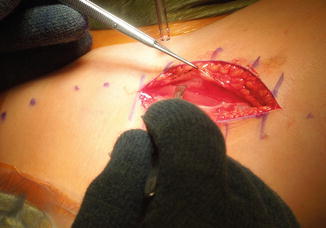
Exposure
A soft tissue protector and retractor may be inserted into the wound (Alexis Wound Protector, Rancho Santa Margarita, CA). Identify the junction of the anterior two-thirds and posterior one-third of the TFL. Make an incision in the fascia over the TFL parallel to its fibers, and this may be extended both distally and proximally beyond the length of the skin incision. The location of the fascial incision over the muscle helps protect the LFCN.
Pick up the anterior flap of the fascia (Fig. 3). Dissect muscle fibers sharply from the fascia or bluntly using finger dissection to delineate the anterior and medial borders of the tensor muscle belly. A medial retractor will retract the rectus femoris. A blunt-tipped retractor can be placed between the superior hip capsule and the gluteus minimus to aid in retracting the TFL. Placement of the retractor directly onto the posterior femoral neck should be avoided in preservation cases. Incise longitudinally the fascia over the lateral rectus, which will facilitate its medial retraction. Split the retinaculum overlying the hip, and the lateral circumflex vessels to the hip are visualized (Fig. 4). At the anterior inferior iliac spine (AIIS), the two heads of the RF can be seen.
 Neuromuscular Hip Disorders: Focus on Cerebral Palsy
 Femoral Deformities: Varus, Valgus, Retroversion, and Anteversion
 Surgical Technique: Arthroscopic Labral Management
 Surgical Technique: Open Proximal Hamstring Repair
 Subspine Impingement and Surgical Technique
 Atraumatic Instability and Surgical Technique


Related posts:
Neuromuscular Hip Disorders: Focus on Cerebral Palsy
Femoral Deformities: Varus, Valgus, Retroversion, and Anteversion
Surgical Technique: Arthroscopic Labral Management
Surgical Technique: Open Proximal Hamstring Repair
Subspine Impingement and Surgical Technique
Atraumatic Instability and Surgical Technique
Stay updated, free articles. Join our Telegram channel
Full access? Get Clinical Tree
