Fig. 10.1
(a, b) Demonstrates vascular architecture of the glenoid labrum, via a sagittal section of a human cadaveric specimen (a). Note the watershed zone in the anterosuperior region. A clear synovial recess (sr) is shown in (b), located between the labrum (L) and glenoid (g) (Adapted from Cooper et al. [1]. Reprinted with permission)
Numerous cadaveric and clinical studies have documented important distinct anatomic variations in both the biceps anchor attachment and the superior labrum, respectively. These variations must be appreciated when attempting to distinguish a “true” SLAP (superior labrum anterior posterior) lesion and further respected when providing appropriate treatment to pathology about the superior labral complex. Roughly 50 % of shoulders will demonstrate the biceps tendon originating from the supraglenoid tubercle, with the remaining half demonstrating its fibers originating directly from the superior labrum [7]. Furthermore, the majority of shoulders display a biceps with a posterior or posterior dominant glenoid labrum insertion, with only a minority of shoulders demonstrating an equal distribution of fibers inserting on the anterior and posterior aspect of the labrum [7, 8]. Grossly, the biceps anchor will usually demonstrate normal physiologic mobility, and overconstraint after repair may be an important factor leading to postoperative stiffness [6].
Normal anatomic variations to the superior labrum have also been reported with differing degrees of incidence, based on arthroscopic findings during surgery. Three distinct variants are classically described that included a sublabral foramen, a sublabral foramen with a cord-like middle glenohumeral ligament (MGHL), and an absent anterosuperior labrum with a cord-like middle glenohumeral ligament (Buford complex) [9, 10]. Aside from a sublabral “hole,” the aforementioned sublabral recess may represent a potential space underneath the superior labrum adjacent to the biceps attachment site – which is also commonly associated with a cord-like MGHL [11]. These variations have been noted to occur in 13 % of the population, with the “Buford complex” being the least common [9]. From a clinical standpoint, these variations have been observed to play a role in the pathogenesis and predilection for lesions about the superior labral complex and influence abnormal biomechanics about the glenohumeral joint. In a large prospective series of 546 patients, Kim and colleagues revealed that the presence of a superior labrum anatomic variant had a positive association with anterosuperior labral fraying, an abnormal superior glenohumeral ligament, and increased passive external rotation with the arm in abduction [12]. Furthermore, both the sublabral foramen variant and the “Buford complex” were shown to have an increase association with type II SLAP lesions [4, 12]. It is critical to recognize and understand the significance of these variants and ultimately distinguish them from pathologic lesions, as errant repair will often result in postoperative pain and stiffness, with an inferior clinical outcome [4].
10.1.2 Classification (Fig. 10.2)

Fig. 10.2
Shows illustrations of the various SLAP lesions, based upon the original classification system (with modifications) (Adapted from Powell et al. [19]. Reprinted with permission)
Since the first description on a superior labral lesion near the biceps tendon origin in an overhead throwing athlete by Andrews and colleagues [13], numerous classifications systems have been developed to aid in the understanding and treatment of these injuries [14–19]. Snyder et al. coined the termed “SLAP” lesion to denote a tear of the superior labrum anterior posterior and reported an incidence of 6 % in over 2,000 shoulder arthroscopies [14, 15]. These authors also developed the most commonly utilized classification system with four distinct types of tears [14].
Type I lesions describe degenerative fraying of the superior labrum’s free edge with an intact and stable biceps anchor. This particular entity is usually the result of age-related degenerative changes and should not necessarily be considered the primary pathology in patients with underlying shoulder pain [20]. Type II tears represent an unstable lesion in which the superior labrum and biceps anchor are detached for the glenoid rim – frequently this complex will be symptomatic and displaced into the glenohumeral joint. These lesions are reported to be the most common subtype, representing 41 % of SLAP tears in Snyder’s original article [14]. A bucket-handle tear of the superior labrum with an intact biceps anchor represents a type III tear. Depending on the size and morphology (meniscoid superior labrum) of the torn labrum, mechanical symptoms may ensue, as the torn fragment will often displace into the joint. Type IV lesions represent a bucket-handle tear of the superior labrum that extends into the biceps anchor. Variable amounts of biceps tendon proper may be involved in the pathology, which may ultimately affect surgical management.
Type II lesions are often described as the most clinically relevant subtype based on their frequency [2, 6], and as a result Morgan and colleagues developed a subclassification system for the type II SLAP tears [17]. These authors proposed tears to be further quantified based on location and extension of the tear, “A” being more anterior, “B” being more posterior, and “C” being a combined anterior posterior lesion. Furthermore, a type IIB lesion may develop posterosuperior instability with glenohumeral “pseudolaxity” [17]. Choi and Kim have also described a type II variant where destabilization of the superior bicipital–labral anchor complex is accompanied by a concomitant articular cartilage avulsion that can lead to loose bodies within the glenohumeral joint [18]. The so-called combined lesions have also been described and classified by Maffet and Powell, respectively [16, 19]. Maffet initially expanded upon the original Snyder classification system, noting that a review of his own patients demonstrated that only 62 % fit within the original schema. He describes type V tears as a Bankart lesion that extended into the superior labrum. Type VI lesions are denoted by a type II tear with an unstable labral flap, and finally type VII lesions represent tears that extend through the MGHL – resulting in an incompetent capsuloligamentous complex [16]. Powell further described types VIII through X, which involve a type II lesion with posterior extension, circumferential extension, or a concomitant posteroinferior labral disruption (reverse Bankart), respectively [19].
More recently, a myriad of studies have tried to enumerate the agreement between observers when diagnosing SLAP lesions, based upon Snyder’s initial criteria, with varying results [21–23]. Gobezie et al. utilized video vignettes to establish inter- and intraobserver agreement for both the diagnosis and treatment of SLAP lesions. Findings of this study demonstrated considerable interobserver variability and only moderate intraobserver variability (κ = 0.54 & κ = 0.45) in regard to both treatment and diagnosis of a SLAP lesion. Furthermore, surgeons had difficulty distinguishing a normal shoulder from type I and II SLAP tears. Interestingly, arthroscopists were more likely to agree on treatment of the lesion, rather than how they would classify it based upon the Snyder criteria [21]. Jia and colleagues, in a similar study, were able to demonstrate improved intraobserver and interobserver agreement of SLAP tear diagnosis and classification (κ = 0.67 and 0.804), among experienced shoulder arthroscopists. Simplifying the labrums into normal or abnormal increased absolute agreement and intraobserver reliability, and utilization of the Morgan subclassification system did not affect the average correlation coefficient. Of note, quality of the video vignettes significantly affected the clinician’s ability to make a confident diagnosis [22]. The lack of surgeons’ ability to physically probe the labrum and perform arthroscopic impingement maneuvers (“peel off” of the labrum) has been cited as an intrinsic limitation of these studies [21, 22].
In a more clinically relevant study, Wolf et al. investigated the influence of multiple patient variables (via clinical vignettes) on the classification and treatment of superior labral complex injuries [23]. The variables included were age, sex, job activity, sports participation, and history/physical examination findings. Surgeons included in the study were part of the MOON (Multicenter Orthopedic Outcomes Network) shoulder group. Based on those surgeons surveyed, age, vocation, sporting activity, and physical examination findings were determined to be the most critical variables to affect treatment choices. These variables resulted in a treatment change 36 % of the time and a Snyder classification system change 28 % of the time [23]. Importantly, it must be noted that all these studies are colored by the fact that universal treatment standards do not exist for the various SLAP pathologies and that age and activity level often significantly influence the treatment algorithm, for the patient and surgeon alike.
10.1.3 Pathogenesis
Numerous etiologies have been proposed for the pathogenesis and underlying shoulder biomechanics responsible for the creation of a SLAP lesion. Frequently accepted mechanisms of injury include forceful traction loads to the arm, direct compression loads, and repetitive overhead throwing activities [6]. Acute traumatic injuries may be responsible for a SLAP tear in a contact athlete – typically the result of a direct blow to the adducted shoulder [24]. Biomechanical studies have demonstrated that impaction loading to a forward flexed arm is more likely to produce an acute SLAP lesion, in comparison to the arm in the extended position [25]. Inferior traction injuries, weightlifters or fall while water skiing, have also been described as clinically and biomechanically culpable for the acute SLAP injury [16].
The overhead athlete usually presents as a distinct and unique patient population, at particular risk for the development of a SLAP lesion. Regardless of the precise mechanisms, lesions are the direct consequence of repetitive overhead throwing activities that occur as the result of overuse. The motions of hyperabduction and external rotation result in an increase in shear and compressive forces on the glenohumeral joint and ultimate strain on the rotator cuff and capsulolabral structures [26]. The dominant arm of young male high-performance overhead athletes appears to be most at vulnerable patient population [27]. The position of the shoulder has been shown to play a role in both biceps stability and injury pattern to superior labrum/biceps anchor. Although some controversy exists as to whether or not late cocking or the deceleration phase of throwing places the superior labrum at risk for injury, the biceps tendon insertion demonstrates 20 % less strength during late cocking [28]. Further biomechanical data that mimics throwing motion was only able to demonstrate increase strain in the superior labrum during the late cocking phase [29].
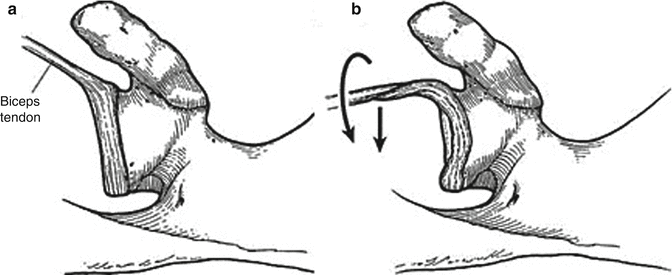
Specific to the throwing athlete, various anatomic and biomechanical factors may result in a predisposition to development of injury patterns in the superior labral complex. These athletes often develop a shift in shoulder range of motion, with an increase in external rotation, which can be accompanied with or without maintenance of total arc range of motion. These motion changes can be associated with bony changes, capsular changes, or both. This underlying phenomenon has been termed “GIRD” (glenohumeral internal rotation deficit) [26, 30, 31]. Wilk and colleagues have demonstrated that pitchers with a diagnosis of GIRD, based on physical examination, have an increased risk of shoulder injury. In this 3-year prospective study of 122 pitchers, those carrying a diagnosis of GIRD were twice as likely to develop a shoulder injury [32]. Numerous biomechanical mechanisms have been postulated to result in SLAP tears of the overhead athletes; Burkhart’s [33] theory of a proposed “peel-back” mechanism is one such etiology (Fig. 10.3) [33]. This theory suggests that the inciting events of posterior and inferior glenohumeral capsular contractures lead to repetitive microtrauma in the overhead athlete, with a relative posterior and superior shift of the humeral head during the cocking phase of throwing. Such a shift in glenohumeral kinematics then marks an increase in shear forces at the posterosuperior labrum. The biceps will then adopt a more vertical position that creates a “vicious cycle” where torsional forces are then generated at the posterosuperior labrum. This recurrent shear and torsional force combination at the bicipital–labral complex will then lead to a “peeling back” of the labrum toward the scapular neck [27, 33]. The sine qua non of such a lesion is the posterior capsular contracture, which must be addressed during the treatment phase with dedicated stretching.


Fig. 10.3
Depicts the “peel-back” mechanism. Resting position of the biceps anchor viewed superiorly (a). The biceps will move posteriorly and twist at its base in the abducted and externally rotated position, resulting in labral “peel back” (b) (Adapted from Burkhart et al. [58]. Reprinted with permission)
A second proposed biomechanical mechanism for the production of a SLAP lesion is that of internal impingement. This theory implies that the superior labrum is subjected to shear and direct contact stresses in the late cocking position of throwing. The SLAP lesion is ultimately a result of impingement of the articular portion of the rotator cuff and posterosuperior labrum between the humerus and glenoid rim [34, 35]. The inciting event, however, appears to be subtle anterior shoulder instability, secondary to muscle fatigue or ligamentous injury. Such instability will allow the humeral head to shift anteriorly during abduction and external rotation (late cocking), and the aforementioned impingement ensues. Such a shift in glenohumeral mechanics has been corroborated in biomechanical studies mimicking anterior capsular laxity and concomitant posterior capsular contracture [36]. Champions of this model emphasize the need for treatment of the labral tear and anteroinferior instability.
A final method for the production of a SLAP injury is that of a “weed-puller” mechanism, initially described by McLeod and Andrews [13]. In this theory torsion produced by the long head of the biceps brachii tears the labrum away from the glenoid. Distinct from the other proposed mechanisms is that this particular theory suggests that the deceleration phase of throwing is the underlying culprit producing the SLAP tear. This theory was first developed on the basis of biomechanical cadaveric data that showed peak biceps muscle activity during the deceleration motion [37].
It is imperative for the clinician to evaluate the athlete as a whole when attempting to discern the underlying etiology of the SLAP lesion. Throwing requires a complex series of coordinated movements that ultimately transmit large amounts of energy from the lower trunk to the arm – the so-called kinetic chain [38]. Alterations in this cascade can result in motions and stresses that injure the labrum. In a similar fashion the role of the scapula and its overall contribution to shoulder motion and the kinetic chain must also be respected. The scapula’s synchronized relationship with the humerus allows for a stable center of rotation of the glenohumeral joint. The overhead thrower can become susceptible to scapular dyskinesis, which may eventually lead to the “SICK” scapula (scapular malposition, inferior medial border prominence, coracoid pain, malposition, and dyskinesis of scapular movement) [39]. This abnormal position of the scapula can lead to abnormal kinematics of the glenohumeral joint and pathologic stress across the labrum, ultimately leading to disability of the throwing shoulder.
10.2 Diagnosis
10.2.1 History
The clinical diagnosis of a SLAP tear can often pose a challenge, even to the most experienced surgeon. Patients will often display concomitant pathology of the shoulder, based on preoperative history, physical exam, and imaging, with symptoms consistent with an insidious onset of nondescript pain. A thorough history, including the mechanism of injury, must be elicited, as the significance of a SLAP lesion, even at the time of surgery, can often be unclear.
Pain is the most common clinical complaint and is usually located anteriorly. Athletes will associate the pain with athletic impairment, including loss of throwing velocity or difficulty with overhead motions [27, 40]. In the overhead athlete mechanical symptoms can predominate, and the sensation of catching, popping, or clicking will be present with rotational movements. Symptoms of weakness and instability may be the result of other underlying pathologies such as partial-thickness rotator cuff tears, capsulolabral injuries, biceps tendinopathy, and internal impingement [41]. Weakness should be carefully evaluated, as it may be the result of a ganglion cyst formation and compression of the suprascapular nerve. Additionally, “dead arm syndrome,” although typically associated multidirectional instability of the glenohumeral joint, has also been described in athletes with SLAP tears [42].
10.2.2 Physical Examination
The physical examination of the athlete, in the face of a potential SLAP lesion, should commence with assessment of both glenohumeral and scapulothoracic motion of the affected shoulder. It is imperative that glenohumeral range of motion be assessed with the scapula stabilized and compared to the contralateral extremity. As previously discussed overhead throwing athletes often exhibit findings consistent with GIRD, defined as a deficit of internal rotation of at least 20° of glenohumeral motion when compared to the contralateral side [26]. As a result, shoulder rotation must be evaluated in adduction and 90° of abduction and should be performed in the supine position to assist with scapular stabilization. If two examiners are available, one can stabilize the scapula by placing a hand over the coracoid and acromion while the other measure the arc of motion. Alternatively, these maneuvers can be successfully performed with a single examiner as well. Judicious evaluation of shoulder stability must also take place, as combined lesions of anterior capsulolabral structures are not uncommon in the overhead athlete. Anterior instability can be assessed, with maintenance of the supine position and utilization of the load shift and apprehension relocation testing. It is also very important to evaluate for possible inferior and posterior instability, with the sulcus sign and posterior apprehension or jerk testing, respectively. Manual rotator cuff strength testing must also be documented, as these muscles function as important dynamic stabilizers of the glenohumeral joint.
Stay updated, free articles. Join our Telegram channel

Full access? Get Clinical Tree


