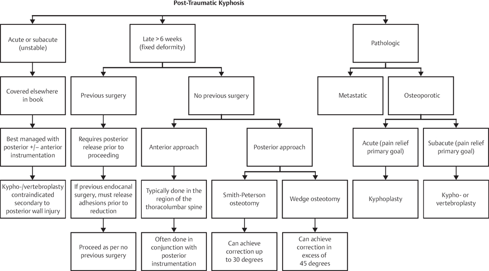
42 Kyphotic deformities of the spinal column may result in overall sagittal imbalance and in turn may cause pain, affect gait, and affect neural structures and adjacent segments of the spine. Although there are no absolute measurements of normal lordosis and kyphosis, general ranges are acceptable. Lumbar lordosis of 60 degrees (40–80) and thoracic kyphosis of 35 degrees (20–50) generally constitute normal sagittal contours. The thoracolumbar junction generally has neutral alignment. Abnormal thoracic kyphosis (over 50 degrees) most commonly is associated with poor posture and has no specific structural pathology. Postural kyphosis carries with it a benign course and responds well to exercises and behavioral modifications. Structural kyphotic deformities, however, may require a more intensive workup and treatment. Scheuermann disease is the most common structural thoracic kyphotic deformity in the juvenile population. Scheuermann described the disease, first reported in 1920, as vertebral wedging with various endplate irregularities seen on radiographs that resulted in the “round back” appearance in the adolescent. Sorensen later described the radiographic pathognomonic findings of the disease to be the anterior wedging of at least 5 degrees in three consecutive thoracic vertebrae.1 Schmorl described the radiographic appearance of intervertebral disk herniations that are often seen in the anterior vertebral endplates in this disease (Schmorl nodules). The etiology of the disease is unclear. Aseptic necrosis of the ring apophysis, collagen composition changes, hereditary factors, endocrinologic imbalance, and others have been proposed. Despite the lack of definitive evidence for its pathogenesis, the presentation and treatment of Scheuermann disease are well described. The two main types of Scheurmann kyphosis are the thoracic (type 1 or most common) and the thoracolumbar (type 2). The thoracolumbar type, although less common, is more frequently associated with curve progression in adulthood. The kyphosis leads to abnormal biomechanical forces on the ring apophysis. Compressive forces anteriorly and tension forces posteriorly during growth are the likely culprits in the progression of the deformity. It is reported to occur in anywhere from 1% to 8% of the population. Scheuermann disease is most commonly diagnosed during adolescence, despite its earlier onset. The most common presenting complaint is a cosmetic deformity or poor posture. Pain associated with the deformity may occur as well, most commonly associated with adult presentation and long-standing kyphosis. Parents will usually complain of their child’s poor posture and round shoulders. As opposed to postural kyphosis, where the deformity is corrected by hyperextension, Scheuermann kyphosis is more rigid. In order for the sagittal balance to stay neutral, lumbar hyperlordosis is often seen in adolescence. Neurologic complaints are exceedingly rare in adolescence but may occur in adulthood when associated with a superimposed degenerative disk herniation, dural cysts, or a severe deformity.2 Familial occurrence is described in the literature and will not uncommonly be described by the patient and family.3 Inspection will often reveal a round back–type deformity that will often worsen with forward bending but not correct itself with hyperextension, as postural kyphosis often will. Generally, no other gross abnormalities will be found, and it is rare to pick up neurologic complaints or findings, but looking for long-tract signs is an important part of the exam. As with scoliosis, general deformity parameters exist where visceral organs may be affected. Clinical respiratory findings are rare and generally do not occur until curves reach greater than 100 degrees. Pulmonary function tests have shown abnormalities at curve magnitudes as low as 80 degrees. Plain radiographs offer most if not all the information needed to make the diagnosis. Standing full-length radiographs are paramount to the diagnosis. Three consecutive wedged vertebrae of 5 degrees or more establish the diagnosis. Several other radiographic findings are associated as well. Schmorl nodules, multilevel early disk degeneration, endplate irregularities, and increased incidence of spondylolysis have all been described in the literature. Flexion and extension radiographs will often show the increased kyphosis on forward bending with the associated lack of correction on hyperextension (at the apex of the deformity). Computed tomography (CT) scans are rarely indicated. Magnetic resonance imaging (MRI), however, may be useful in cases of neurologic complaints or preoperatively. Nonoperative approach is the mainstay of treatment for curves under 70 degrees. Exercises, medications, and activity modification have all been associated with symptom improvement. Bracing has played a role in the treatment of Scheuermann kyphosis in the growing child. Well-molded thoracolumbosacral orthoses (TLSOs) have shown some improvement in the overall curve magnitude. The efficacy of brace treatment, however, is debatable, and the hard indications for it are unclear. The treating physician must weigh the cons of brace treatment in the adolescent versus the potential pros when recommending this treatment. Operative fixation is generally recommended for curves over 90 degrees. At curves of this magnitude and greater, pain, pulmonary dysfunction, and positive sagittal balance may respond only to surgical intervention. Curves between 70 and 90 degrees generate the most debate regarding surgical treatment. Multiple factors may play a role in the decision making. Progressive deformity, cosmesis, pain, and neurologic deficits may all play a role in surgical intervention at curves of this magnitude. Different surgical approaches have been described over the years in the literature. With the evolution of spinal instrumentation, the need for anterior-posterior surgery has decreased. Generally, a posterior-only approach using Smith-Petersen osteotomies (see Chapter 44) and segmental fixation account for the vast majority of surgical interventions today. Brace treatment in the skeletally immature has yielded inconsistent results. With the use of braces, correction of up to 50% may be expected; however, several studies report loss of correction at follow-up and variable outcomes.4 Progression despite bracing is also commonly reported. Surgical intervention offers a definitive solution for severe cases and deformities that continue to be symptomatic despite nonoperative treatment. Anterior and posterior approaches were commonly used in the past for correction of the deformity. Comparative studies have shown that the outcomes following a posterior-only surgery are similar to anterior-posterior techniques. With higher complications described from the anterior-posterior approach, a posterior-only approach is most commonly used today. Surgical correction of Scheuermann kyphosis generally entails a posterior-only-based correction. Using multiple segmental fixation and Smith-Petersen osteotomies, deformity correction upwards of 50% can be well maintained and minimal loss of correction can be expected. Numerous surgical complications have been described: hemothorax, pneumothorax, infection, and paraplegia have all been reported. Avoiding the anterior approach has decreased pulmonary complications. Neurologic injury is a potentially catastrophic complication and has been reported as often as 1 in 700–1000 surgeries. Both motor and sensory evoked potentials are routinely monitored intraoperatively to give the surgeon real-time evaluation of spinal cord function. The most common mechanism that is thought to be responsible for spinal cord injury is the stretch of the anterior spinal artery during the correction. Immediate release of the correction should follow any loss of neural function. Given the high sagittal junctional stresses following kyphosis correction, junctional kyphosis and/or screw (or hook) pullout have been reported. Anchoring the construct to the first lordotic level distally is important. Other surgical complications reported are similar to those reported for other deformity/scoliosis surgeries and are not unique to Scheuermann disease. 2. Ogilvie JW, Sherman J. Spondylolysis in Scheuermann’s disease. Spine 1987;12(3): 251–253 PubMed 3. Damborg F, Engell V, Andersen M, Kyvik KO, Thomsen K. Prevalence, concordance, and heritability of Scheuermann kyphosis based on a study of twins. J Bone Joint Surg Am 2006; 88(10):2133–2136 PubMed 4. Lowe TG, Line BG. Evidence based medicine: analysis of Scheuermann kyphosis. Spine 2007;32(19, Suppl):S115–S119 PubMed Coe JD, Smith JS, Berven S, et al. Complications of spinal fusion for Scheuermann kyphosis: a report of the Scoliosis Research Society Morbidity and Mortality Committee. Spine 2010; 35(1):99–103 PubMed The authors reviewed a series of 683 patients undergoing spinal fusion for Scheuermann kyphosis. They reported a complication rate of 14%, including infection (3.8%), acute neuroligcal injury (1.9%), spinal cord injury (0.6%), and death (0.6%). Complications were more common in older patients. There was no difference in complications among anterior, posterior, or combined approaches. Denis F, Sun EC, Winter RB. Incidence and risk factors for proximal and distal junctional kyphosis following surgical treatment for Scheuermann kyphosis: minimum five-year follow-up. Spine 2009;34(20):E729–E734 PubMed The authors followed 67 patients for 5 years to determine the incidence of proximal junctional kyphosis (PJK) and distal junctional kyphosis (DJK). PJK was observed in 20 patients (30%) and was thought be associated with disruption of the ligamentum flavum or failure to incorporate the proximal end vertebrae. DJK was seen in 8 patients (12%) and was thought to be related to fusions falling short of the first lordotic disk. Geck MJ, Macagno A, Ponte A, Shufflebarger HL. The Ponte procedure: posterior only treatment of Scheuermann’s kyphosis using segmental posterior shortening and pedicle screw instrumentation. J Spinal Disord Tech 2007;20(8):586–593 PubMed The authors describe the technique for correcting Scheuermann kyphosis using Ponte osteotomies and pedicle screw instrumentation. They report successful use of the technique in 17 patients with results equivalent to combined anterior-posterior techniques and with minimal loss of correction over 5-year follow-up. Damborg F, Engell V, Andersen M, Kyvik KO, Thomsen K. Prevalence, concordance, and heritability of Scheuermann kyphosis based on a study of twins. J Bone Joint Surg Am 2006;88(10): 2133–2136 PubMed The authors analysed a cohort of almost 35,000 twins from the Odense-based Danish Twin Registry. They found an overall prevalence of 2.8%, 3.6% among men and 2.1% among women. A strong genetic link was seen in monozygotic twins, with heritability noted to be 74%.
Scheuermann Disease
![]() Classification
Classification
![]() Workup
Workup
History
Physical Examination
Imaging Studies
![]() Treatment
Treatment
![]() Outcomes
Outcomes
![]() Complications
Complications
References
Suggested Readings

Stay updated, free articles. Join our Telegram channel

Full access? Get Clinical Tree


