Fig. 8.1
“Medial pulley transosseous-equivalent” repair depicting a modified TOE construct with a broad medial inter-implant mattress configuration. This construct only requires two tendon suture passes, without compromising biomechanical performance compared to the original TOE construct (which requires a minimum of four tendon suture passes). Technical efficiency is therefore improved (Adapted from Park et al. [27])
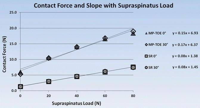
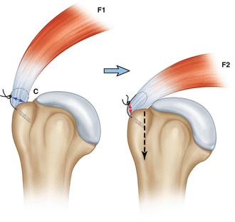
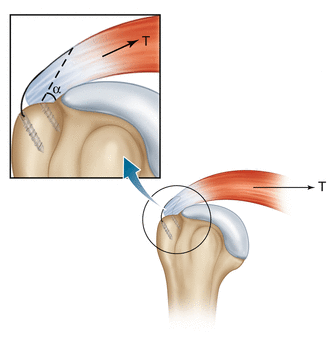
Tendon suture-bridging constructs have been theorized to be “self-reinforcing” in the face of potentially destructive tensile loading forces, mimicking a Chinese finger trap where distraction forces also contribute to resistance to separation [3]. This concept has been biomechanically characterized and validated [26]. Force sensors were placed on the footprint prior to repair with the MP-TOE technique in ten cadaveric specimens. With progressive tendon loading, the frictional forces increased as well. Using the same methodology, a standard “single-row” (SR) repair demonstrated the same relationship; however, the progression of resistive frictional force was disproportionately more with the MP-TOE repair (Fig. 8.2). The SR repair creates a “focal loop wedge” effect, where the focal lateral suture loop elongates creating obligatory tendon compression over the lateral footprint with tendon force simulation (Fig. 8.3). With SR repair, tendon loading creates suture loop elongation and coincidental construct failure with increasing gap formation. In this context, SR repair cannot be optimally self-reinforcing as it does not fix the tendon-bridging suture loop medially whereby a more complete footprint “wedge” effect [3] can occur to include the medial footprint (Fig. 8.4).


Fig. 8.2
Footprint contact force with progressive tendon loading. At each load, the MP-TOE repair provided significantly more contact force (p < 0.05) compared to single-row (SR) repair. With increasing loads, the MP-TOE repair had a significantly higher progression (slope) of footprint contact force compared with single-row repair at both 0° (p = 0.025) and 30° (p = 0.014) abduction. If the slopes were not significantly different between repairs, the “self-reinforcement” effect would not have been validated

Fig. 8.3
Schematic rendering showing the “focal loop wedge” effect. With increasing tendon load (F1 to F2), focal loop stitch configurations (as seen with single-row repairs) elongate, thus compressing the focal loop stitch (blue to red double arrows), while exposed footprint contact area C is relatively decreased. In turn, the F2 load is creating a focal compression vector (down arrow) onto the footprint. This is an example of a rotator cuff “self-reinforcement” effect. However, this effect is significantly improved with tendon-spanning constructs that are fixed medially to secure a “loop” that can bridge the entire footprint; this has been shown to provide disproportionately more progressive footprint frictional resistance even with disruptive tendon loading forces [26]

Fig. 8.4
Medial fixation secures a “loop” of suture that spans the footprint, allowing for a more complete footprint restoration, which increases the compressive vector over the insertion even with tendon loading. As the load T increases, the angle decreases, wedging the tendon between the tendon-bridging sutures and bony insertion (“wedge” effect). With single-row repair, the suture loop is not fixed medially, as the tendon is secured over the isolated anchors only (From Burkhart et al. [3])
Rotator cuff tear classification for full-thickness tears can be based most intuitively on footprint anatomy: Stage 0 or normal, Stage I supraspinatus only, Stage II overlap area (supraspinatus and infraspinatus), Stage III the anterior half of the infraspinatus, and Stage IV the entire supraspinatus and infraspinatus tendons [17]. While this classification is based on functional anatomy, intraoperative technical decision-making is based on tendon mobility and tissue quality (tendon and bone).
8.3 Clinical Presentation and Essential Physical Examination
The athlete with a rotator cuff injury will typically present with either an acute traumatic injury or a chronic history of more than 6 months having had persistent and progressive pain. The tendon regions commonly involved include the supraspinatus tendon and anterior aspect of the infraspinatus tendon. For an acute injury, the mechanism of injury may point to the type and extent of injury. For chronic injuries, the athlete can usually be participating in sports that involve repetitive overhead motion, such as baseball, volleyball, tennis, water polo, and the like. The prognosis for acute injury after repair is arguably more predictable than for chronic injury with a repetitive overuse etiology insofar as return to sport in the latter setting involves recreating the very mechanism that contributed to the threshold injury. Furthermore, truly chronic tears may be associated with muscle atrophy and fatty infiltration that may be irreversible. Each patient may present different functional goals after surgery largely based on the level of play (e.g., “weekend warrior” to professional), and this will frame the reasonable expectations that can be achieved after repair.
A careful physical examination begins with inspection. The affected extremity may manifest atrophy; and gross atrophy within the scapular fossae could be diagnostic for a rotator cuff tear—a spinoglenoid notch cyst with suprascapular nerve compression would be in the differential diagnoses, however, and careful examination for labral pathology must be considered particularly in the athlete who performs repetitive exertional overhead motion. Passive range of motion is critical to assess to eliminate a concurrent diagnosis of frozen shoulder or glenohumeral osteoarthritis. Excessive external rotation relative to the normal shoulder may signify a torn subscapularis tendon. Active range of motion testing generally will gauge the functional limitations the patient may have.
Motor testing can isolate the muscles involved. Resisted forward elevation in the scapular plane can help assess supraspinatus involvement, although a negative test does not mean a tear is not present, especially when the tear is only partial full-thickness, leaving a tear that is functional on examination. In addition to elevation, the supraspinatus assists rotation of the humerus internally and externally. Weakness to external rotation at the side generally means the patient has supraspinatus and infraspinatus involvement—the patient may elevate the elbow to compensate. However, a negative test does not preclude a supraspinatus tear, even though the tear may be full-thickness as it may be only partially torn in the anterior-posterior dimension. The belly-press, lift-off, and bear-hug tests can help measure subscapularis tendon pathology, although this tendon is not commonly injured relative to the supraspinatus and infraspinatus tendons. The belly-press test is performed with the hand pressing maximally into the abdomen and the elbow in line with the trunk in the sagittal plane—a positive test is manifested by a relative weakness compared to the normal side or the elbow dropping posteriorly in the sagittal plane. The lift-off test is performed by placing the dorsum of the hand against the mid-lumbar spine—a positive test is apparent when the patient is unable to internally rotate and lift the hand away from the back. The bear-hug test is performed with the hand placed on the contralateral shoulder over the acromioclavicular joint—a positive test results when the examiner can externally rotate the hand away from the initial position.
Neer and Hawkins tests are typically used to assess external impingement underneath the coracoacromial ligament arch. Pain posteriorly with abduction and external rotation, with or without instability, indicates the possibility of internal impingement, particularly in an overhead throwing athlete, for example. Other provocative tests can be used to assess for non-tendinous concurrent pathologies, including labral tears, which can dictate the type of imaging study that is obtained, and the surgical approach.
8.4 Essential Radiology
Radiographic x-ray imaging can provide baseline information regarding the athlete’s shoulder. When rotator cuff pathology is suspected in the athlete, true anterior-posterior, outlet, and axillary views are routinely obtained. Arthritic changes of the glenohumeral and acromioclavicular joints will give a measure of relative overuse. Glenohumeral osteoarthritic changes would lend to a poor prognosis with respect to rotator cuff tendon pathology. In a more senior athlete, a high-riding humeral head would also be a poor prognostic factor, consistent with chronic overuse, suggesting a large to massive rotator cuff tear involving both the supraspinatus and infraspinatus tendons. The acromiohumeral interval (AHI), the distance between the acromion and humerus, has been inversely related to rotator cuff tearing and fatty infiltration [33]. Fatty infiltration can be classified by using computed tomography (CT). Grade 0 has been defined as normal muscle, grade 1 fatty streaking, grade 2 more muscle than fatty infiltration, grade 3 equal amounts of muscle and fat, and grade 4 more fatty infiltration than muscle. Increasing grade of infiltration has been correlated with worsening function [4, 11, 12], and high grades have been associated with irreversible changes, even after repair.
Magnetic resonance imaging, in addition to gauging fatty infiltration, can directly assess the degree of tendon involvement, and a geometric classification has been proposed to aid in predicting repair potential and prognosis after repair [7] (Fig. 8.5); care should be taken to identify the anterior cord of the supraspinatus tendon. T2-weighted coronal and sagittal oblique views should be evaluated. In the type 1 tear, the tear is retracted on the coronal view more than it is wide on the sagittal view, with retraction <2 cm; this would be equivalent to a crescent-type tear, with a relatively good to excellent prognosis. The type 2 tear is more retracted than it is wide, with the width being <2 cm; this is equivalent to a longitudinal tear (“L” or “U”) also with a good to excellent prognosis. Massive contracted tears would define the type 3 tear with retraction and width being greater than or equal to 2 cm, and the prognosis is more guarded. Cuff tear arthropathy defines the type 4 “tear” using this classification. This geometric classification allows for recommendations on repair technique based on the classification type: Type 1 would allow for end-to-bone repair, type 2 margin convergence, type 3 interval slides or partial repair, and type 4 arthroplasty.
8.5 Disease-Specific Clinical and Arthroscopic Pathology
The spectrum of injury involving the athlete’s rotator cuff includes tendonitis, partial tears (bursal- and articular-sided), and full-thickness tears. The athlete with a torn rotator cuff will typically have pain that prevents full participation; sport-specific activity will usually be limited with respect to range of motion and strength. Arm activity above shoulder level is particularly affected. Athletes with chronic tears may participate until threshold injury occurs, which may involve some degree of tear progression. Either the athlete will cycle back to a rest-and-participation cycle or threshold injury prevents return.
During arthroscopy, the diagnosis can be confirmed. A spectrum of injury may be encountered from tendonitis and interstitial tearing, to partial tearing, to full-thickness tears. In tears that occur from acute injury, pathology may be isolated to the rotator cuff. Acute tears can arise when the force between the greater tuberosity and acromion displaces the rotator cuff, usually seen more in the contact athlete. Either partial- or full-thickness tears may occur. Chronic tears may manifest structural degeneration of the coracoacromial ligament (external impingement), in which case acromioplasty should be carefully considered [35].
In the overhead throwing athlete, the labrum and biceps tendon must be carefully inspected and characterized. Adaptive changes may give false-positive findings, and care must be taken not to aggressively treat all findings beyond simple debridement; otherwise, there may be an increased risk for over-constraining the shoulder. This may lead to a return to sport, but at a lower level, or possibly be career-ending [21]. Internal impingement from overuse throwing can manifest as partial articular-sided tears of the infraspinatus tendon and partial tearing of the posterosuperior labrum.
8.6 Treatment Options
A spectrum of pathology may be present in the athlete, whether arising from an acute or chronic setting: tendonitis and interstitial tearing, to partial bursal- or articular-sided tearing, to full-thickness tearing. In general, the smaller or lesser the extent of injury, the more likely nonoperative management may be effective. And acute trauma may have a better prognosis for both nonoperative and operative management, relative to chronic overuse injuries. For overuse injuries, return to sport can be difficult when the sport activity itself contributes to the injury.
In general, all types of rotator cuff injuries can be considered for nonoperative management. Perhaps most importantly, a requisite healing period must be respected, to give the biology of healing an opportunity. Range of motion must be maintained. Typically passive and active range of motion should be progressed in a graded manner; in the throwing athlete, attention toward stretching the posterior capsule may be necessary. Routine nonsteroidal anti-inflammatory medications and icing may be initiated. A cortisone-type injection trial can be considered, with strict counseling to avoid a premature return to play and the risk of relative overuse. Once pain is controlled, progressive conditioning may be initiated. Generally, rotator cuff injuries can take 3–6 weeks minimum prior to return to sport; injuries that involve loss of structural integrity usually take longer, up to 12 weeks minimum. Each patient’s progress must be individualized, and attention should be given to the timing (symptom duration), the sport-specific requirements, and the patient’s goals. Although individualizing treatments is recommended, generally, full-thickness tears in the athlete warrant surgical intervention or strong consideration with a relatively lower threshold to perform surgery, as the natural history is for tears to progress [39].
When nonoperative management fails, arthroscopic surgery can be considered. For tendonitis or tears involving less than 50 % of the tendon substance, including either bursal- or articular-sided tears, careful debridement and subacromial decompression can be considered. However, there remains some controversy regarding the indications for acromioplasty; generally when coracoacromial ligament changes exist, acromioplasty should be strongly considered. For partial articular-sided tears involving more than 50 % of the footprint, in situ trans-tendon repair and completing the tear followed by repair have been described [14]—discussion here is reserved for the following chapter 9. For bursal-sided tears involving more than 50 % of the medial-lateral substance, including full-thickness tears, a repair can be considered. For small 10 mm tears without retraction, single-anchor repair can be sufficient. For larger tears, SR and TOE repairs can be considered with tissue loss and tendon retraction being important factors in repair strategy decision-making.
8.7 Author’s Preferred Treatment
The lateral decubitus position is preferred with 10 lbs of arm traction and approximately 45° arm abduction; simultaneously, 5 lbs of traction is placed at approximately 30° abduction via a second pulley to allow another option for footprint exposure and implant approach as needed. A standard posterior portal is used to access the glenohumeral joint. Under direct visualization, an anterior portal is made at the rotator interval using needle localization; this is cheated slightly superior to allow for potential anchor placement through this portal as needed. A nerve hook probe is used to perform routine diagnostic arthroscopy with a 30° arthroscope; relatively rarely, a 70° arthroscope can be used for subscapularis tears. In the athlete, attention must be given to the capsulolabral structures including the biceps tendon. The rotator cuff tear can be thoroughly characterized by careful inspection and probing, with both intra- and extra-articular arthroscopy. After switching the posterior cannula to the subacromial space, an anterolateral working portal can be established after needle localization; care must be taken to not place this portal too superior or inferior, accounting for access and exposure with respect to suture passing and distal-lateral anchor placement. If a kissing lesion on the coracoacromial ligament is encountered (usually seen in athletes with a chronic history), the ligament is released with a heat probe; acromioplasty when indicated is performed at the end of the case to limit bleeding and risk for adversely affecting visualization during the repair. A posterolateral portal, also established with needle localization, is often used to achieve a bird’s-eye view of the torn tendon.
Stay updated, free articles. Join our Telegram channel

Full access? Get Clinical Tree


