, Carina Cohen2 and Katherine C. Faust1
(1)
Department of Orthopaedics, Tulane University, New Orleans, LA, USA
(2)
Ortopedia e Traumatolgia, Cirurgia de Ombroe e Cotovelo, UNIFESP Hospital Israelita Albert Einstein, São Paulo, Brazil
Keywords
Rotator interval Rotator cable Coracohumeral ligament Interval closure Superoglenohumeral ligament 11.1 Introduction
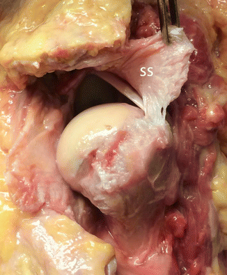
The rotator interval is the triangular part of the glenohumeral joint between the anterior supraspinatus and superior subscapularis muscle borders, extending medially from the coracoid process and laterally the transverse humeral ligament which connects the greater and lesser tuberosities (Fig. 11.1). The term rotator interval originated from the landmark 1970 paper by Neer classifying proximal humerus fractures; he described it as a “ligamentous area between the tendons of the supraspiantus and subscapularis” [1, 2]. The superior and middle glenohumeral ligaments travel across the interval, along with the long head of the biceps tendon and the. The father of shoulder surgery, E. A. Codman, wrote that “The coraco-humeral and gleno-humeral ligaments should never have been described as entities,” describing them as variable portions of the glenohumeral capsule [3]. However, with the advent of arthroscopic surgery and improved magnetic resonance imaging techniques, these portions of the rotator interval have proven consistent and useful anatomic landmarks.
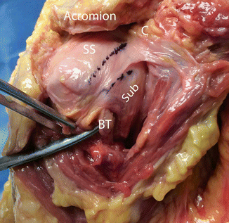

Fig. 11.1
Right shoulder from anterolateral with ink marks outlining the borders of the supraspinatus (SS) and subscapularis (Sub). The lateral and anterior portions of the deltoid have been detached exposing the anterior part of the humerus. The rotator interval extends from the coracoid (c) to lay over the long head of the biceps tendon (BT), which has been exposed in its groove
11.2 Rotator Interval Gross Anatomy
During dissection of the glenohumeral joint, the rotator interval is found between the supraspinatus and subscapularis muscles, whose fibers contribute to the interval [4]. The ligamentous tissue making it up consists of the superior glenohumeral ligament, arising from the supraglenoid tubercle and inserting on the greater tuberosity, and the, discussed in the next section, along with glenohumeral capsular tissue. These ligaments are distinct and blend with the bursal and deep fascia of the glenohumeral joint (Jost et al. [4]) (Fig. 11.2). The superior glenohumeral ligament runs deep to the coracohumeral ligament and can be better seen from inside the joint, while the coracohumeral ligament is better seen from outside (Figs. 11.3 and 11.4). The structure becomes loose with humeral head elevation and internal rotation, taut with humeral head depression and external rotation.
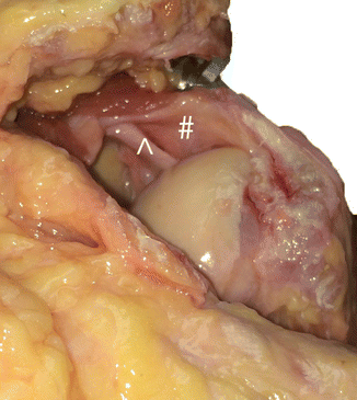
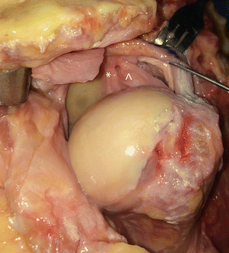
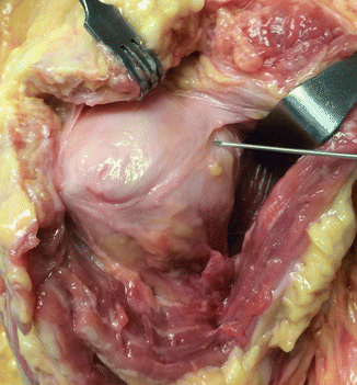

Fig. 11.2
Right shoulder, viewed from posterior to anterior after reflection of the infraspinatus and teres minor, to reveal the (#) and superior glenohumeral ligament (^), distinct entities that resist external rotation and inferior translation of the humeral head

Fig. 11.3
Right shoulder looking from posterior to anterior after reflection of the infraspinatus and teres minor to allow visualization of the rotator interval. The superior glenohumeral ligament (^) can be seen extending toward the long head of the biceps tendon as it enters its groove. The middle glenohumeral ligament (*) can also be seen crossing the rotator interval from this view

Fig. 11.4
Right shoulder with army-navy retractor subcoracoid and Senn rake around the acromion looking from anterior to posterior. The needle points out the broad as it travels from the coracoid to the greater and lesser tuberosities
11.3 Coracohumeral Ligament Gross Anatomy
The has been described in Cunningham’s and Morris’ anatomy textbooks since their early editions as a ligament arising from the root and lateral border of the coracoid process, along with the conjoint tendon, and inserting on the greater tubercle [5, 6]. It consists of two bands, one which forms the anterior sling of the bicipital groove and invests in the subscapularis fascia and the other which forms the posterior sling and invests the supraspinatus fascia [7] (Figs. 11.5 and 11.6). Studies have shown that this anterior portion inserts on the lesser tuberosity [4]. It is this bicipital groove portion of the coracohumeral ligament that blends with the transverse humeral ligament to form the biceps pulley. The coracohumeral ligament also splits to incorporate both the superficial fascia of the muscles on their bursal side and the deep glenohumeral capsule [8, 9] (Fig. 11.7). The deep portion of the posterior sling thickens to form the that runs perpendicular to the supraspinatus and infraspinatus fibers. Morris’ Human Anatomy describes the ligament from posterior as an “uninterrupted continuation of the capsule” and from anterior as “a fan-shaped prolongation” [5]. The ligament becomes taut with external rotation and lax with internal rotation of the humeral head (Fig. 11.8).

