Revision Total Knee Arthroplasty: Component Removal and Modular Selection
Patient Presentation and Symptoms
Total knee arthroplasty has become a very common orthopedic procedure. Excellent results have been obtained with proper technique, good instrumentation, and correct indications. Failure of knee arthroplasty can occur for a variety of reasons, including infection, component loosening, and instability. Patients present with pain, limited motion, and swelling.
Indications
- Infection
- Aseptic loosening
- Osteolysis secondary to polyethylene wear
- Instability
Contraindications
- Incompetent extensor mechanism
- Infection with a highly virulent organism
- Excessively ill patient who is not a surgical candidate
Physical Examination
- May be unable to bear full weight on the extremity
- Knee effusion is common.
- Knee malalignment
- Range of motion may be restricted
- Check knee stability
- Check the skin for warmth and erythema.
Diagnostic Tests
- Radiographs are mandatory. In fact, an effort should be made to obtain x-rays taken prior to the original arthroplasty, after the original arthroplasty, and any taken up to the present time.
- Computed tomography (CT) scan may a useful adjunctive test to check for areas of occult osteolysis.
- Bone scan and indium scans can be helpful in diagnosing infection and aseptic loosening.
- Aspiration with fluid analysis should be done to rule out infection.
Preoperative Planning and Timing of Surgery
Once the cause of failure has been identified, surgery should be scheduled. If the knee is infected, treatment should be undertaken as soon as possible. For aseptic failure, surgery can be performed electively; however, for cases with gross component loosening and osteolysis it is preferred to undertake the revision as soon as feasible. The reason for this urgency is to avoid greater bone loss. In the preoperative planning, the amount of bone loss should be estimated so that the appropriate implant and modular augments are available. It may be necessary to have a structural allograft available, such as a femoral head, distal femur, or proximal tibia.
Special Instruments
- Standard total knee arthroplasty instrumentation
- Modular revision arthroplasty instrumentation and components, including femoral and tibial augments, as well as stem extensions
- Full set of flexible osteotomes, curettes, and rongeurs
- Small sagittal saw for component removal
- Slap-hammer extraction tool
Anesthesia
Regional anesthesia is recommended. However, this decision should be made collectively by the patient, the surgeon, and the anesthesiologist after discussing the options and obtaining the patient’s informed consent.
Patient Position
The patient should be positioned supine with a tourniquet on the upper thigh.
Surgical Procedure
- Every effort is made to use the old skin incision, which can be extended proximally and distally as needed. A standard medial parapatellar arthrotomy is preferred because it can be converted easily to a quadriceps snip if necessary for better exposure.
- Aspiration of synovial fluid upon entering the joint. This is sent for cell count, stat Gram stain, and culture; synovial tissue is sent for a frozen section.
- Complete synovectomy with reestablishment of the medial and lateral gutters
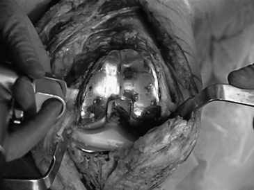
- Component removal: Following removal of the modular tibial polyethylene articulation, the femoral component is removed. A small sagittal saw is used to interrupt the component cement interface (Fig. 40–1). Alternatively, you could use a Gigli saw. A flexible osteotome is then passed around the implant to further loosen it from the femur. This is a very effective technique to remove the component without removing excess bone. This technique can also be used to remove a cementless component. The tibia and patella are removed in similar fashion.
- Debris removal: We used small curettes and rongeurs to removal all debris and soft tissue from the bone surfaces. Once this is done, adequate assessment of bone loss can be performed (Fig. 40–2).
- Bone loss classification: Bone loss is classified as symmetric or asymmetric and contained versus uncontained (Fig. 40–3).
- The goal of revision total knee arthroplasty (TKA) is to create equal flexion and extension gaps. When this is not readily achieved, adjustments need to be made. It is important to remember that adjustments on the femoral side can affect the knee in either flexion or extension, whereas any adjustments on the tibial side will affect both.
- The surgical technique involves re-creating the femur, including size and rotation; rebuilding the flexion space on a flat tibial surface; and reestablishing the extension space
- Management of bone loss: Tibia: In general, small contained defects greater than 5 mm can be filled with cement, whereas larger contained defects less than 5 mm may be filled with impacted cancellous bone allograft. Larger uncontained defects, less than 5 mm, usually require modular augmentation or even structural allograft (Fig. 40–4). Femur: Femoral defects are usually uncontained and can be managed with modular augments. Large defects that extend proximal to the epicondyles usually require structural allografts.
- Once the flexion and extension gaps are balanced, and the appropriate alignment and stability have been confirmed with provisional implants, the final components are assembled. In preparation for cementing, the bony surfaces are cleaned with pulsatile lavage. Cement is placed around the core implant, with care taken to avoid cement around the stem extensions. The stem extensions are inserted in a cementless press-fit manner.
- The tourniquet is let down and hemostasis is obtained.
- Closure is performed with interrupted No. 0 absorbable sutures to close the arthrotomy. A Hemovac drain is left deep in this layer. The subcutaneous layer is closed with 2-0 absorbable sutures. The skin is closed with staples and the wound is dressed with a light sterile dressings.

Stay updated, free articles. Join our Telegram channel

Full access? Get Clinical Tree


