14 Ligament Injuries: Anterior Cruciate Ligament Reconstruction with Hamstring Tendons
Patient Presentation and Symptoms
Anterior cruciate ligament (ACL) tears are among the most common injuries seen by the orthopedic surgeon. Patients with ACL tears may report feeling or hearing a pop in the knee during the injury. As the ACL is a relatively vascular ligament, many patients also develop a significant effusion/hemarthrosis within 1 or 2 hours of the injury. Patients may complain of give-way episodes. Those with significant pain may also have concomitant meniscal or articular cartilage pathology.
Indications
Anterior cruciate ligament reconstruction, substituting a free tendon graft for the torn native ligament, is a common surgical procedure. Because the menisci, articular surfaces, and other restraining structures around the knee are susceptible to injury during episodes of instability, it is generally accepted that ACL reconstruction should be offered to patients who have or are at risk of having recurrent knee instability.
The goal of ACL reconstruction is to restore normal anterior knee stability, and when deciding on surgical intervention, the orthopedist has to decide which graft substitute best accomplishes this goal. The four-stranded semitendinosus/gracilis (ST/G) graft has many advantages over other grafts, including its strength, stiffness, and cross-sectional area. A four-stranded ST/G graft looped over a femoral cross pin and secured on the tibia with the IntraFix tibial fastener (Mitek, Norwood, Massachusetts) is a strong and stiff graft construct, and easily allows for unrestricted and aggressive rehabilitation without the risks of graft elongation or failure. We currently recommend this graft-fixation construct for ACL reconstruction.
Contraindications
Absolute contraindications for ACL reconstruction include active infections and limited knee range of motion. While debatable, nonsurgical treatment is offered to patients who do not experience knee instability, those not involved in sports that require cutting and jolting, and those who are willing to modify their lifestyles to avoid knee instability. Also debatable is the treatment of the adolescent patient with open growth plates. Due to concerns for growth arrest as a result of transphyseal drilling, most surgeons recommend conservative treatment if there is more than 1 cm of growth remaining.
Physical Examination
The examination of the patient with a suspected ACL injury must be sufficiently thorough to assess for concomitant injuries. Specific tests for ACL injuries include the Lachman test and the pivot-shift test. The Lachman test gives the examiner two vital pieces of information: the amount of anterior tibial translation and the quality of the end point to the ACL.
Diagnostic Tests
Every patient with a suspected ACL tear should have radiographs of the knee. The radiographs in the acute setting should be viewed for fractures, avulsion fractures, and osteochondral injuries. Radiographs in the chronic ACL-deficient patient may include spurring of the inter-condylar eminences, intercondylar notch narrowing, depression of the sulcus terminalis, and osteophyte formation of the inferior patella and medial compartment. While magnetic resonance imaging (MRI) is sensitive and specific in diagnosing ACL tears, it should be rarely needed to do so. An MRI scan, however, can give the clinician additional information on the extent of bone bruising, meniscal pathology, and other ligamentous injuries that may affect treatment decisions.
Another diagnostic test that can aid in the diagnosis of an ACL tear is the arthrometer. Although many different arthrometers exist, we have used the KT-1000 (MedMetric Corporation, San Diego, California) exclusively. Side-to-side differences of 3 mm between the affected and unaffected knee with a manual maximum force are diagnostic of an ACL tear.
Special Considerations
Surgeons can consider extraphyseal reconstructions using the semitendinosus and gracilis tendons in adolescents with open growth plates. Other considerations include the use of osteotomies in the ACL-deficient patients with malalignment and arthrosis.
Preoperative Planning and Timing of Surgery
The surgeon should review the radiographs and, if obtained, the MRI of the knee prior to surgery. The surgeon should be prepared to perform concomitant procedures such as meniscus repair and should be able to adequately assess and treat articular cartilage pathology. While debatable, most surgeons will postpone elective ACL reconstruction until knee range of motion is restored following an acute injury. If the knee motion is limited as a result of a locked, bucket-handle meniscus tear, one option is to repair the meniscus and postpone the ACL reconstruction until range of motion is restored.
Special Instruments
The surgeon can use a variety of special instruments to help with the reconstruction. In addition to basic arthroscopy instrumentation, the surgeon can use tibial and femoral tunnel guides, tibial tunnel dilators to compact the tibial metaphyseal bone, drill bits of various sizes to drill the tibial and femoral bone tunnels, tendon strippers to harvest the semitendinosus and gracilis tendons, and manufacturer-specific instrumentation for graft fixation in the femur and tibia.
Anesthesia
Anterior cruciate ligament reconstruction is now routinely performed as an outpatient procedure. The choice of anesthesia type is up to the patient. Options include general anesthesia, spinal or epidural anesthesia, and regional nerve blocks. In some cases, a regional block can be added to the main anesthesia choice to aid in postoperative pain management.
Patient and Equipment Positions
The patient is placed in the supine position on a well-padded table. A tourniquet is applied to the proximal thigh but is not routinely used unless needed for hemostasis and visualization during arthroscopy. A lateral post is used as a fulcrum when applying a valgus load to the knee to visualize the medial joint space.
Surgical Procedure
Graft Harvest
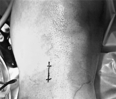
Figure 14-1 Photograph of the 1.5-inch tendon harvest incision on the proximal tibia, ~2 cm medial to the tibial tubercle.
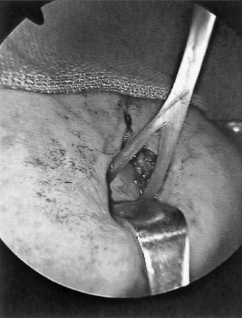
The semitendinosus and gracilis tendons are harvested through a 1.5-inch incision centered ~2 cm medial to the tibial tubercle (Fig. 14–1). Dissection is carried down to the sartorial fascia, which is incised parallel and distal to the palpable semitendinosus tendon. The semitendinosus and gracilis are then released from their tibia attachment, and each tendon is whip-stitched with nonabsorbable suture. The tendons are freed from surrounding adventitia using blunt dissection. Also, extratendinous bands are incised to completely free up the tendon to their respective sheaths (Fig. 14–2). It is important to incise these fas-cial bands to prevent premature amputation of the tendon short of its muscle belly. The tendons are then harvested with a blunt-end tendon stripper and taken to the back table, where they are prepared by removing attached muscle and the free ends are whip-stitched with No. 2 braided polyester suture. The graft is sized in preparation for tunnel drilling.
Related posts:

Stay updated, free articles. Join our Telegram channel

Full access? Get Clinical Tree


