CHAPTER 4 Plica Synovialis of the Elbow
Documentation of synovial plica as an anatomic structure began in 1912, when Testut described the “humeral radialis labrum.”1,2 Descriptions of this as a pathologic condition began in the 1950s.3–5 Since then, the pathologic condition has been described as the lateral synovial fringe,6 snapping plica,7 and synovial fold of the humeral radial joint.1 Identification of the plica as a pathologic condition can be traced to individual case reports within the past 30 years that described symptoms, clinical presentation, and treatment. The true significance has been dramatically enhanced with the advent of arthroscopy.
ANATOMY
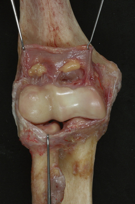
Most of the early and recent anatomic descriptions of this structure come from the Japanese or French literature.8–12 One of the most detailed accounts was provided by Isogai and colleagues.10 Anterior and posterior folds have been documented in embryo and adult elbows, confirming these folds as normal anatomic structures (Fig. 4-1).13 The so-called lateral fold has been identified only in adults.10 This finding prompted the theory that a single event, repetitive trauma, or anatomic variant combined with activity may give rise to the development of the pathologic structure. This theory is enhanced by recognition that the histology of the normal anterior and posterior folds is that of fibrous fatty tissue; however, the lateral plica consists of hyaluronized bundles with fibrous tissue, making it histologically different from the anterior and posterior bands.10 Others have recognized that histologically, normal plica reveals uninflamed fibrofatty synovial tissue with no evidence of necrosis.1 However, a hard, fibrous type of tissue is characteristic of the lateral or circumferential variation of the plica.
Duparc and coworkers1 performed careful dissections on 50 elbows and demonstrated some form of a synovial fold in 43 (86%). They described six orientations, including four (9%) that were circumferential. In 30, the structure was described as rigid, and in the remaining 13, it was soft and pliable. The relationship with degenerative changes of the radial head was also recognized. A later German investigation sought to define the accuracy of MRI detection of the plica. These investigators determined that MRI detected some form of a plica in 88 studies. Dividing the folds into small (31%), medium (57%), and large (12%), they also correlated degenerative changes with the larger plicae.14
PATIENT EVALUATION
Early recognition of the potential pathologic features of this tissue has been offered in case reports.9 Circumferential synovial flow was found to roll over the radial head and then slide back over the radial neck with extension and flexion, respectively.6 Duparc and collagues1 also recognized that 4 of 43 folds were actually circumferential and seemed to blend with the annular ligament and the margin of the radial head. It is this particular variant that appears to be the pathologic lesion (see Fig. 4-1).
Clinical Presentation
In the early descriptions, the pain was confused with that of tennis elbow. It might have been confusion with this lesion that prompted the so-called Bosworth approach to tennis elbow.15 In this procedure, arthrotomy and inspection of the joint was recommended because it was recognized that intra-articular changes correlated to lateral elbow pain. Numerous investigators have subsequently pointed out the difficulty of differentiating a pathologic plica from lateral epicondylitis when the characteristic snapping is absent.1,6,16 The diagnosis becomes simplified when the clinical presentation is not so much lateral or posterolateral joint pain, but rather catching, snapping, and locking. The differential diagnosis may be tennis elbow or a loose body, depending on the presence or absence of the mechanical features.6,7

Stay updated, free articles. Join our Telegram channel

Full access? Get Clinical Tree


